Pathophysiology of Cardiac Arrhythmias: Cardiac arrhythmias are abnormalities in the rate, rhythm, or electrical conduction of the heart. Under normal conditions, electrical impulses originate from the sinoatrial (SA) node, which acts as the natural pacemaker of the heart. These impulses spread through the atria, pass through the atrioventricular (AV) node, and travel through the bundle of His, bundle branches, and Purkinje fibres to produce coordinated contraction of the heart.
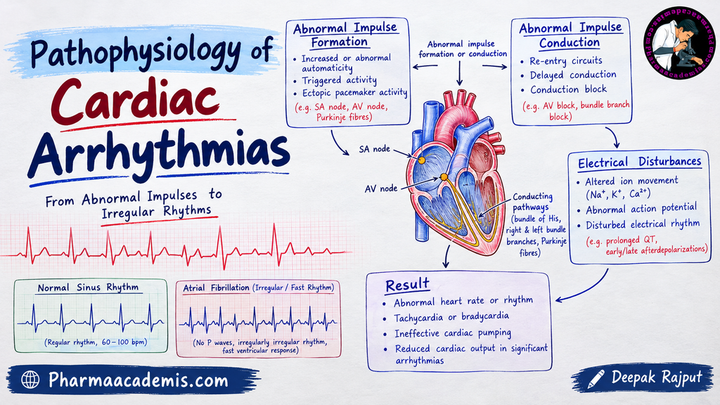
When the generation or conduction of electrical impulses becomes abnormal, the heart may beat too fast (tachycardia), too slowly (bradycardia), or irregularly. These abnormalities can reduce the efficiency of blood circulation and may lead to symptoms such as palpitations, dizziness, breathlessness, and fainting. Severe arrhythmias may cause heart failure or sudden cardiac death.
Pathophysiology of Cardiac Arrhythmias
The pathophysiology of cardiac arrhythmias mainly involves abnormalities in impulse formation, impulse conduction, or a combination of both. These disturbances affect the normal electrical activity of the heart and alter its rhythm.
1. Abnormal Impulse Formation
Normally, the SA node generates electrical impulses at a regular rate and controls the heart rhythm. However, abnormalities in pacemaker activity or the development of impulses from other cardiac tissues can produce arrhythmias.
Increased automaticity: Automaticity is the ability of cardiac cells to generate electrical impulses spontaneously. When automaticity increases, cardiac cells may generate impulses more rapidly than normal, resulting in tachycardia or premature heartbeats. This may occur due to sympathetic stimulation, myocardial ischaemia, or certain electrolyte disturbances.
Decreased automaticity: When the SA node generates impulses more slowly than normal, the heart rate decreases. This may occur due to increased vagal activity, ageing-related changes in the conduction system, or certain medications. In severe cases, it may result in bradycardia or pauses in the heartbeat.
Ectopic impulse formation: Sometimes, electrical impulses originate from locations other than the SA node, such as the atria or ventricles. These abnormal impulses may produce premature atrial or ventricular contractions and may occasionally initiate more sustained arrhythmias.
2. Triggered Activity
Triggered activity occurs when abnormal electrical depolarizations develop during or after a cardiac action potential and produce additional impulses. It is commonly associated with disturbances in ion movement or calcium regulation within cardiac cells.
It is classified into two types:
Early afterdepolarizations (EADs): These occur before repolarization is complete, usually when the cardiac action potential is prolonged. They may trigger abnormal ventricular rhythms, particularly torsades de pointes, which is associated with QT interval prolongation.
Delayed afterdepolarizations (DADs): These occur after repolarization has been completed. They are commonly associated with excessive calcium accumulation inside cardiac cells, which can trigger additional electrical impulses. Digoxin toxicity and excessive catecholamine stimulation are possible causes.
Both types of triggered activity can produce premature heartbeats and may contribute to tachyarrhythmias.
3. Abnormal Impulse Conduction
Electrical impulses normally travel through the cardiac conduction system in a coordinated sequence. When conduction is delayed or interrupted, the normal rhythm of the heart may be disturbed.
Conduction delay: Electrical impulses travel more slowly than normal through a part of the conduction system. This may interfere with coordinated cardiac activation and contribute to arrhythmias.
Conduction block: Electrical impulses may be delayed excessively or fail to pass through a particular region. For example, an atrioventricular (AV) block interferes with the transmission of impulses from the atria to the ventricles. Depending on its severity, it may cause a slow heart rate, dizziness, or fainting.
Conduction abnormalities may develop due to myocardial infarction, fibrosis, inflammation, electrolyte disturbances, or medications that affect electrical conduction.
4. Re-entry Mechanism
Re-entry is one of the most important mechanisms responsible for cardiac tachyarrhythmias. Normally, an electrical impulse activates cardiac tissue once and then stops because the tissue temporarily becomes refractory to further stimulation.
In re-entry, an electrical impulse travels through an abnormal circuit and returns to tissue that has recovered its ability to respond. The impulse can then reactivate the tissue and continue circulating repeatedly, producing a rapid heart rhythm.
This mechanism usually requires a suitable conduction pathway, differences in conduction or recovery between tissues, and the ability of the returning impulse to stimulate previously activated tissue.
Re-entry is involved in atrioventricular nodal re-entrant tachycardia (AVNRT), atrial flutter, and many forms of ventricular tachycardia.
5. Electrolyte Imbalance and Ion Channel Abnormalities
The electrical activity of cardiac cells depends on the movement of sodium (Na⁺), potassium (K⁺), and calcium (Ca²⁺) ions across the cell membrane. These ions are essential for depolarization, repolarization, and normal impulse conduction.
Abnormal electrolyte concentrations can disturb these processes and increase the risk of arrhythmias.
Hypokalaemia (low potassium levels) may alter repolarization and increase susceptibility to certain arrhythmias. Hyperkalaemia (high potassium levels), particularly when severe, can impair conduction and produce serious cardiac disturbances. Abnormal calcium regulation may cause changes in contraction and trigger additional electrical impulses.
These disturbances may occur due to kidney disease, dehydration, certain medications, or other medical conditions.
6. Myocardial Ischaemia and Structural Heart Disease
Myocardial ischaemia occurs when the heart muscle does not receive sufficient oxygen to meet its metabolic requirements. The resulting reduction in energy production and changes in ion movement can disturb the electrical properties of cardiac cells.
Ischaemia may slow conduction, alter repolarization, and increase electrical instability, potentially producing premature beats or serious ventricular arrhythmias.
Conditions such as myocardial infarction, cardiomyopathy, and cardiac fibrosis can also damage the normal electrical pathways. Scar tissue may interfere with impulse conduction and create circuits that support re-entry. Therefore, patients with structural heart disease have an increased risk of developing potentially dangerous arrhythmias.
7. Role of the Autonomic Nervous System
The autonomic nervous system regulates heart rate and electrical conduction through sympathetic and parasympathetic activity.
Sympathetic stimulation: The sympathetic nervous system increases heart rate, accelerates AV nodal conduction, and enhances cardiac excitability. Excessive stimulation during stress, exercise, or certain pathological conditions may promote tachycardia and abnormal impulse formation.
Parasympathetic stimulation: Parasympathetic activity, mainly through the vagus nerve, slows SA node firing and AV nodal conduction. Excessive vagal activity may cause bradycardia or contribute to AV conduction disturbances.
An imbalance between sympathetic and parasympathetic activity can therefore influence the development of cardiac arrhythmias.
Effects of Cardiac Arrhythmias
Cardiac arrhythmias can interfere with the normal pumping action of the heart. When the heart beats too rapidly, the ventricles have less time to fill with blood. When the heart beats too slowly, the amount of blood pumped per minute may decrease.
Some arrhythmias, such as atrial fibrillation, reduce the effectiveness of atrial contraction and may increase the risk of blood clot formation and stroke. Severe ventricular arrhythmias can prevent effective ventricular contraction, causing a sudden fall in blood pressure and cardiac output.
Common symptoms include palpitations, fatigue, dizziness, breathlessness, and fainting. Severe cases may result in heart failure, circulatory collapse, or sudden cardiac death.
Conclusion
Cardiac arrhythmias develop due to abnormalities in impulse formation, impulse conduction, or both. The major mechanisms include abnormal automaticity, triggered activity, re-entry, conduction block, electrolyte disturbances, myocardial ischaemia, and autonomic imbalance. These abnormalities disturb the normal cardiac rhythm and may reduce the efficiency of blood circulation. Understanding these mechanisms helps explain the development, clinical manifestations, and potential complications of cardiac arrhythmias.
Editorial Note
This article has been carefully researched and written by Deepak Rajput with a focus on accuracy, clarity, and evidence-based healthcare information. Pharmaacademias.com