
Pathophysiology of Cataract: Cataract is a condition in which the normally transparent crystalline lens of the eye becomes progressively opaque, resulting in reduced transmission of light to the retina and causing blurred or decreased vision. Cataract develops because of structural and biochemical changes in lens proteins, particularly crystallins, along with oxidative stress, disruption of lens homeostasis, and aggregation of proteins.
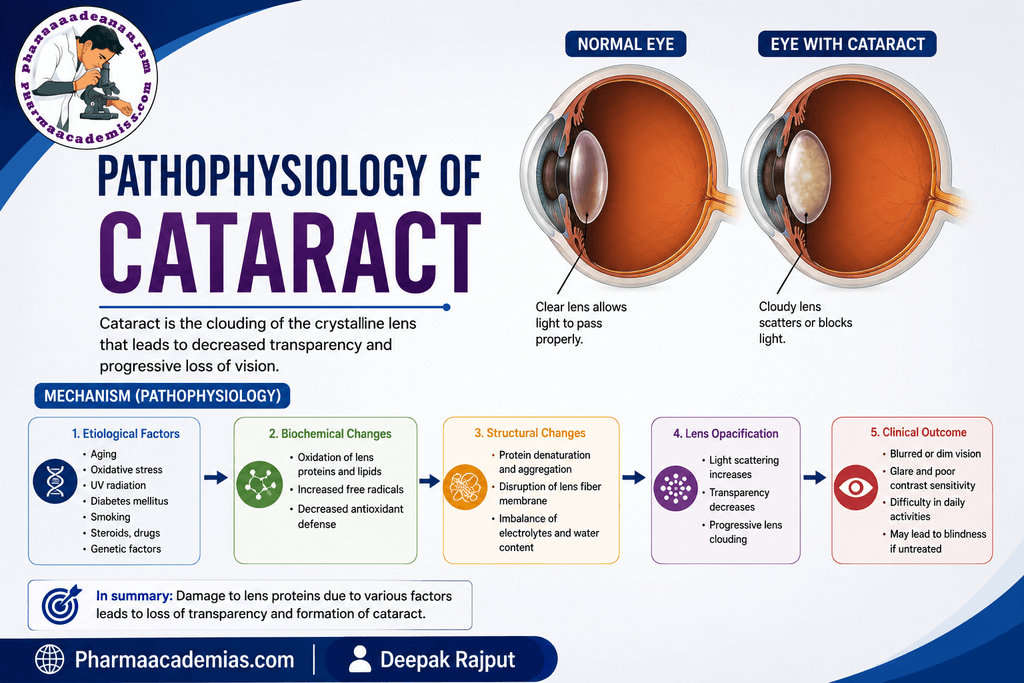
Step 1: Normal Lens Structure and Function
The normal lens is a transparent, avascular, biconvex structure located behind the iris and pupil.
Its main functions are to:
- Transmit light to the retina.
- Refract light.
- Change its shape during accommodation to focus on objects at different distances.
The lens contains:
Lens capsule → Lens epithelium → Lens fibers → Crystallin proteins
Lens transparency depends on the highly organized arrangement of lens fibers and crystallin proteins.
Step 2: Disturbance of Lens Homeostasis
The lens normally maintains transparency through a carefully controlled balance of:
- Water and electrolyte concentration
- Protein organization
- Antioxidant activity
- Energy metabolism
- Ion transport
- Protein repair and degradation
With aging, metabolic disease, trauma, radiation, certain drugs, or other factors, these mechanisms may become impaired.
Step 3: Oxidative Stress
One of the most important mechanisms involved in cataract formation is oxidative stress.
The lens is exposed to reactive oxygen species (ROS). Normally, antioxidant systems such as:
- Glutathione
- Superoxide dismutase
- Catalase
- Other antioxidant mechanisms
help protect the lens.
With aging or other risk factors, antioxidant defenses may become inadequate.
Step 4: Oxidation and Damage to Lens Proteins
The lens contains high concentrations of crystallin proteins, which are essential for maintaining transparency.
Oxidative stress can modify these proteins.
Protein damage may cause:
- Oxidation of sulfhydryl groups
- Protein cross-linking
- Protein aggregation
- Changes in protein structure
- Reduced protein solubility
The normally soluble lens proteins can therefore become increasingly insoluble.
Step 5: Protein Aggregation
As crystallin proteins become damaged and aggregate, they form larger protein complexes.
These aggregates interfere with the normal passage of light through the lens.
Instead of allowing light to pass through efficiently, the abnormal protein structures scatter incoming light.
Step 6: Changes in Lens Fiber Cells
Lens fibers are highly specialized cells that are arranged in an organized manner.
During aging and cataract formation, lens fibers may undergo:
- Cellular degeneration
- Membrane damage
- Loss of normal organization
- Changes in hydration
- Protein accumulation
These structural changes further reduce the transparency of the lens.
Step 7: Disturbance of Electrolyte and Water Balance
The lens maintains its transparency partly through controlled movement of ions and water.
In cataract formation, changes in membrane permeability and ion transport can disturb the normal balance of:
- Sodium (Na⁺)
- Potassium (K⁺)
- Calcium (Ca²⁺)
- Water
Excessive sodium and calcium accumulation can contribute to cellular and protein damage.
Water movement may also increase, causing lens fiber swelling in some forms of cataract.
Step 8: Calcium Accumulation and Protease Activation
In certain cataracts, increased intracellular calcium can activate calcium-dependent proteolytic enzymes called calpains.
These enzymes can break down structural proteins and contribute to further disruption of lens organization.
Step 9: Lens Hydration and Osmotic Changes
Changes in lens metabolism can alter osmotic balance.
For example, in diabetes, increased glucose availability can increase conversion of glucose to sorbitol through the polyol pathway.
Sorbitol accumulates because it crosses cell membranes relatively poorly.
This causes:
Step 10: Formation of Lens Opacity
The combined effects of:
- Protein aggregation
- Oxidative damage
- Lens fiber degeneration
- Electrolyte imbalance
- Hydration changes
- Structural disorganization
cause the normally transparent lens to become increasingly opaque.
The opacity may initially affect only a small region of the lens.
Over time, it can become more extensive.
Step 11: Reduced Transmission of Light
A healthy lens allows light to pass through and focus on the retina.
Step 12: Changes in Visual Function
As the cataract progresses, several visual symptoms may develop.
Blurred or cloudy vision: The most common symptom is gradual reduction in visual clarity.
Glare: Light scattering within the opaque lens can cause increased glare, particularly from:
- Vehicle headlights
- Bright sunlight
- Strong artificial lights
Halos around lights: Some patients may notice halos around bright light sources.
Reduced contrast sensitivity: The ability to distinguish subtle differences between light and dark areas may decrease.
Color changes: Colors may appear less bright or more yellowish because the aging lens increasingly absorbs shorter-wavelength light.
Refractive changes: Changes in the lens can alter its refractive properties and may temporarily increase myopia (nearsightedness) in some patients.
Step 13: Progressive Visual Impairment
Cataract usually develops gradually.
The sequence is:
Lens biochemical damage
↓
Protein aggregation and fiber changes
↓
Lens opacity
↓
Increased light scattering
↓
Reduced retinal image quality
↓
Blurred vision
↓
Progressive visual impairment
Unlike glaucoma, cataract primarily involves opacity of the lens, rather than primary damage to the optic nerve.
Major Types of Cataract and Their Pathophysiology
1. Nuclear Cataract
This affects the central nucleus of the lens.
With aging, the lens nucleus becomes progressively harder and may become yellow or brown because of accumulation of pigments and biochemical changes.
2. Cortical Cataract
This involves the lens cortex. Changes in lens hydration and electrolyte balance can produce characteristic wedge-shaped or spoke-like opacities extending toward the center of the lens.
These opacities interfere with light transmission.
3. Posterior Subcapsular Cataract
This develops near the posterior surface of the lens, beneath the lens capsule. It can interfere significantly with vision because of its location near the visual axis.
It may be associated with factors such as:
- Corticosteroid exposure
- Diabetes
- Radiation
- Aging
Cataract vs. Glaucoma: Key Difference
| Feature | Cataract | Glaucoma |
| Main structure affected | Lens | Optic nerve/retinal ganglion cells |
| Main pathological change | Lens opacity | Progressive optic neuropathy |
| Common association | Aging, oxidative stress | IOP and optic nerve susceptibility |
| Vision loss | Mainly due to reduced/blurred light transmission | Mainly due to visual field loss |
| Reversibility | Cataract-related opacity can be treated surgically | Established optic nerve damage is generally irreversible |
One-Line Summary
Cataract develops when aging or other risk factors cause oxidative, metabolic, protein, and structural changes in the crystalline lens, leading to protein aggregation and lens opacity, increased light scattering, and progressive impairment of vision.
Editorial Note
This article has been carefully researched and written by Deepak Rajput with a focus on accuracy, clarity, and evidence-based healthcare information. Pharmaacademias.com