The Peripheral Nervous System (PNS) is the part of the nervous system that lies outside the brain and spinal cord. It forms an important communication network between the central nervous system (CNS) and the rest of the body.
The PNS carries sensory information from receptors in the body to the CNS and transmits motor commands from the CNS to muscles and glands. It therefore plays a major role in sensation, movement, coordination, and regulation of internal body functions.
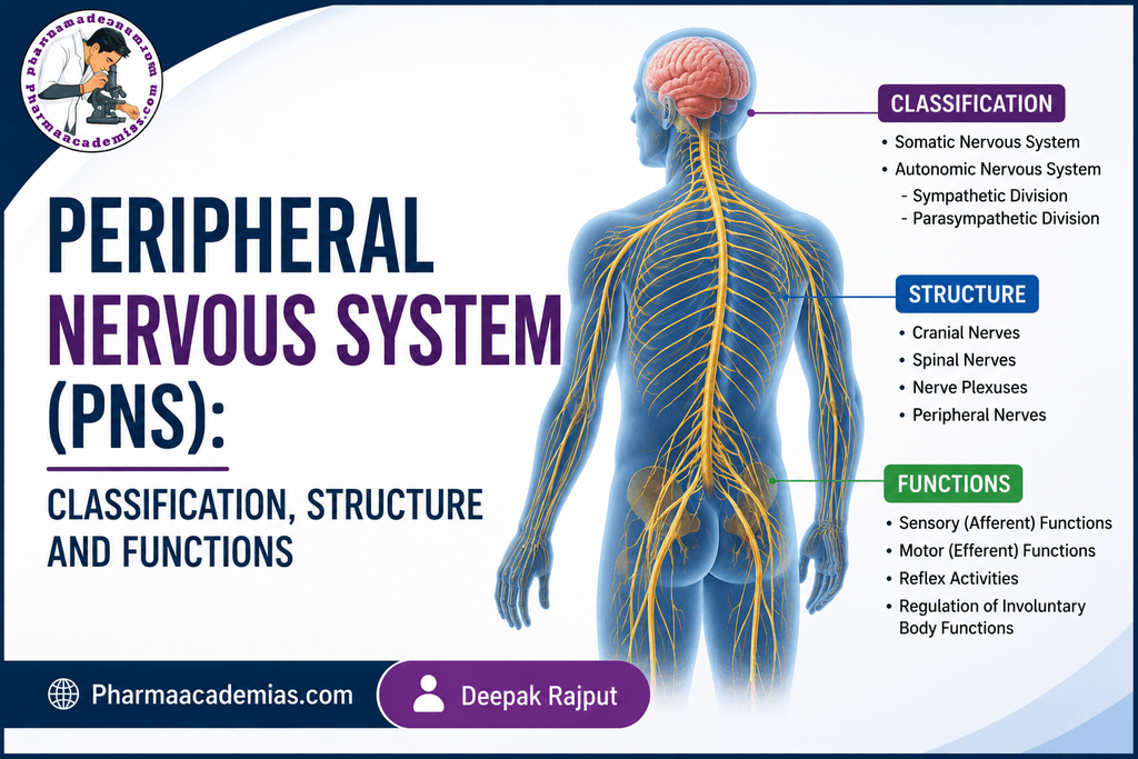
The peripheral nervous system consists mainly of cranial nerves, spinal nerves, peripheral nerves, ganglia, and sensory receptors.
1. Classification of the Peripheral Nervous System
Functionally, the PNS can be divided into two major parts:
A. Sensory (Afferent) Division
The sensory division carries information from the body toward the CNS.
It receives information from:
- Skin
- Muscles
- Joints
- Internal organs
- Special sensory organs
Examples of sensory information include:
- Touch
- Pain
- Temperature
- Pressure
- Stretch
- Body position
- Internal organ sensations
The sensory division can be further classified into:
1. Somatic sensory system:
Carries sensations from the skin, skeletal muscles, joints, and other structures associated with the body wall.
2. Visceral sensory system:
Carries information from internal organs such as the heart, stomach, intestines, and blood vessels.
B. Motor (Efferent) Division
The motor division carries instructions from the CNS to muscles and glands.
It is divided into:
- Somatic nervous system
- Autonomic nervous system
Somatic Nervous System
The somatic nervous system controls skeletal muscles.
It is mainly responsible for voluntary movements such as:
- Walking
- Writing
- Running
- Lifting objects
- Moving the arms and legs
However, some skeletal muscle activities can also occur through reflexes without conscious control.
Autonomic Nervous System
The Autonomic Nervous System (ANS) regulates involuntary functions of the body.
It controls:
- Heart rate
- Blood pressure
- Digestion
- Respiratory airway diameter
- Pupil size
- Sweating
- Urination
- Glandular secretions
The autonomic nervous system is mainly divided into:
- Sympathetic nervous system
- Parasympathetic nervous system
The enteric nervous system is also closely associated with the autonomic nervous system and regulates many functions of the gastrointestinal tract.
2. Autonomic Nervous System
The autonomic nervous system maintains the body’s internal environment (homeostasis). Unlike the somatic system, most autonomic activities occur without conscious control.
For example, when a person is resting, the heart continues to beat, the intestines continue to move food, and glands continue to secrete substances without conscious effort.
The two major divisions have generally opposite but complementary effects:
| Feature | Sympathetic | Parasympathetic |
| General role | Fight or flight | Rest and digest |
| Main activity | Prepares body for activity/stress | Conserves energy and promotes maintenance |
| Heart rate | Increases | Decreases |
| Pupils | Dilates | Constricts |
| Bronchi | Dilates | Constricts |
| Gastrointestinal activity | Generally decreases | Generally increases |
| Salivation | Reduced/less watery | Increased |
| Urinary bladder | Promotes storage | Promotes emptying |
| Energy use | Increases | Conserves energy |
3. Sympathetic Nervous System
The sympathetic nervous system (SNS) is a division of the autonomic nervous system that prepares the body to respond to stress, danger, physical activity, or emergency situations.
It is commonly described as the “fight-or-flight” system.
When a person experiences fear, stress, excitement, or strenuous exercise, sympathetic activity increases.
Structure of the Sympathetic Nervous System
The sympathetic nervous system has a thoracolumbar origin.
Its preganglionic neurons arise mainly from the thoracic and upper lumbar regions of the spinal cord (approximately T1–L2).
The pathway generally contains two neurons:
Preganglionic neuron → Sympathetic ganglion → Postganglionic neuron → Effector organ
Sympathetic Ganglia
Sympathetic ganglia are mainly located close to the spinal cord and include:
- Paravertebral ganglia – arranged as sympathetic chains on either side of the vertebral column.
- Prevertebral (collateral) ganglia – located anterior to the vertebral column, particularly around major abdominal arteries.
The sympathetic system can therefore distribute signals rapidly to several organs at the same time.
Neurotransmitters of the Sympathetic System
At most sympathetic synapses:
- Preganglionic neurons release acetylcholine (ACh).
- Most postganglionic neurons release norepinephrine (noradrenaline).
An important exception is the sweat glands, where sympathetic postganglionic neurons generally release acetylcholine.
The adrenal medulla is another special case. Sympathetic preganglionic fibers stimulate the adrenal medulla, causing it to release mainly epinephrine (adrenaline) and some norepinephrine into the bloodstream.
4. Functions of the Sympathetic Nervous System
The sympathetic nervous system produces several physiological effects.
1. Heart
It:
- Increases heart rate
- Increases force of cardiac contraction
- Increases cardiac output
This helps supply more blood to tissues during physical activity or stress.
2. Blood Vessels
Sympathetic stimulation can cause:
- Vasoconstriction in many blood vessels
- Redistribution of blood flow
- Increase in blood pressure in many situations
3. Eyes
It causes pupil dilation (mydriasis).
This allows more light to enter the eye.
4. Respiratory System
It causes bronchodilation, which increases airflow into the lungs.
5. Gastrointestinal Tract
It generally:
- Decreases gastrointestinal motility
- Decreases digestive activity
- Reduces gastrointestinal blood flow
- Reduces some digestive secretions
During an emergency, digestion becomes a lower priority.
6. Urinary System
It promotes urinary storage by relaxing the bladder wall and increasing contraction of the internal urinary sphincter.
7. Skin
It stimulates:
- Sweating
- Piloerection (goosebumps)
8. Metabolism
It promotes the availability of energy by increasing processes such as:
- Glycogen breakdown
- Fat breakdown
- Glucose availability
9. Adrenal Medulla
Sympathetic stimulation causes the adrenal medulla to release epinephrine and norepinephrine, reinforcing the body’s stress response.
5. Parasympathetic Nervous System
The parasympathetic nervous system (PNS) is the division of the autonomic nervous system that promotes rest, digestion, energy conservation, and routine maintenance of the body.
It is commonly called the “rest-and-digest” system.
It is particularly active when the body is relaxed and after eating.
Structure of the Parasympathetic Nervous System
The parasympathetic nervous system has a craniosacral origin.
Its preganglionic neurons arise from:
- Brainstem nuclei associated with cranial nerves III, VII, IX, and X
- Sacral spinal cord segments, mainly S2–S4
Cranial Component
Parasympathetic fibers travel through:
- Oculomotor nerve (CN III)
- Facial nerve (CN VII)
- Glossopharyngeal nerve (CN IX)
- Vagus nerve (CN X)
The vagus nerve is especially important because it supplies parasympathetic fibers to many organs in the thorax and abdomen.
Sacral Component
Sacral parasympathetic fibers arise from S2–S4 and supply structures including portions of the:
- Large intestine
- Urinary bladder
- Reproductive organs
6. Parasympathetic Ganglia
Unlike sympathetic ganglia, most parasympathetic ganglia are located close to or within the target organs.
Therefore, parasympathetic pathways typically have:
Long preganglionic fiber → Ganglion near/in organ → Short postganglionic fiber → Effector organ
This arrangement allows relatively localized effects.
7. Neurotransmitters of the Parasympathetic System
Both neurons in the parasympathetic pathway primarily use acetylcholine (ACh):
Preganglionic neuron → ACh → Ganglion → Postganglionic neuron → ACh → Target organ
The postganglionic acetylcholine usually acts on muscarinic receptors present on target tissues.
8. Functions of the Parasympathetic Nervous System
1. Heart
It:
- Decreases heart rate
- Reduces cardiac activity, particularly at the atria
This helps conserve energy during rest.
2. Eyes
It causes:
- Pupil constriction (miosis)
- Accommodation of the lens for near vision
3. Salivary Glands
It increases salivary secretion, producing relatively abundant and watery saliva.
4. Gastrointestinal Tract
It promotes:
- Gastrointestinal motility
- Digestive secretions
- Movement of food through the gastrointestinal tract
It therefore plays an important role in digestion.
5. Respiratory System
It generally causes bronchoconstriction and increases bronchial secretions.
6. Urinary System
It promotes:
- Contraction of the urinary bladder
- Relaxation of the internal urinary sphincter
This facilitates urination.
7. Gastrointestinal and Other Glands
It generally promotes glandular secretion and supports normal digestive processes.
8. Sexual Function
Parasympathetic activity contributes to erection, whereas sympathetic pathways are more strongly associated with ejaculation.
9. Sympathetic vs Parasympathetic Nervous System
| Organ/Function | Sympathetic Effect | Parasympathetic Effect |
| Eye – pupil | Dilates | Constricts |
| Heart | Increases heart rate and force | Decreases heart rate |
| Bronchi | Dilates | Constricts |
| Salivary glands | Decreases/changes secretion | Increases secretion |
| Stomach & intestine | Decreases motility and secretion | Increases motility and secretion |
| Blood vessels | Mainly causes vasoconstriction in many vascular beds | Limited direct effect on most blood vessels |
| Urinary bladder | Promotes storage | Promotes urination |
| Sweat glands | Increases sweating | Little/no major direct effect |
| Adrenal medulla | Stimulates secretion of catecholamines | No major direct effect |
| Sexual function | Ejaculation | Erection |
| Overall role | Fight or flight | Rest and digest |
10. Anatomical Differences
| Feature | Sympathetic | Parasympathetic |
| Origin | Thoracolumbar | Craniosacral |
| Spinal origin | T1–L2 | S2–S4 |
| Cranial nerves | Not a major origin | CN III, VII, IX, X |
| Ganglia | Near spinal cord | Near or within target organs |
| Preganglionic fibers | Usually short | Usually long |
| Postganglionic fibers | Usually long | Usually short |
| Main neurotransmitter at ganglia | Acetylcholine | Acetylcholine |
| Main postganglionic neurotransmitter | Usually norepinephrine | Acetylcholine |
| Main function | Emergency/stress response | Rest, digestion and conservation |
Summary
The Peripheral Nervous System connects the central nervous system with the rest of the body. Functionally, it is divided into sensory (afferent) and motor (efferent) divisions. The motor division includes the somatic nervous system and autonomic nervous system.
The autonomic nervous system is mainly divided into the sympathetic and parasympathetic systems. The sympathetic system has a thoracolumbar origin (T1–L2) and prepares the body for stressful or demanding situations through the fight-or-flight response. The parasympathetic system has a craniosacral origin, including cranial nerves III, VII, IX, and X and sacral segments S2–S4, and supports rest, digestion, energy conservation, and routine maintenance.
Together, these systems regulate vital functions such as heart rate, blood pressure, breathing, digestion, pupil size, urination, and glandular secretion, helping maintain the body’s internal balance.
Editorial Note
This article has been carefully researched and written by Deepak Rajput with a focus on accuracy, clarity, and evidence-based healthcare information. Pharmaacademias.com