Pathophysiology of Iron Deficiency Anemia: Iron deficiency anemia (IDA) is the most common nutritional deficiency disorder and one of the most common causes of anemia worldwide. It develops when the body’s iron requirement exceeds iron availability for a prolonged period, resulting in depletion of iron stores, impaired hemoglobin synthesis, reduced red blood cell production, and ultimately decreased oxygen-carrying capacity of blood.
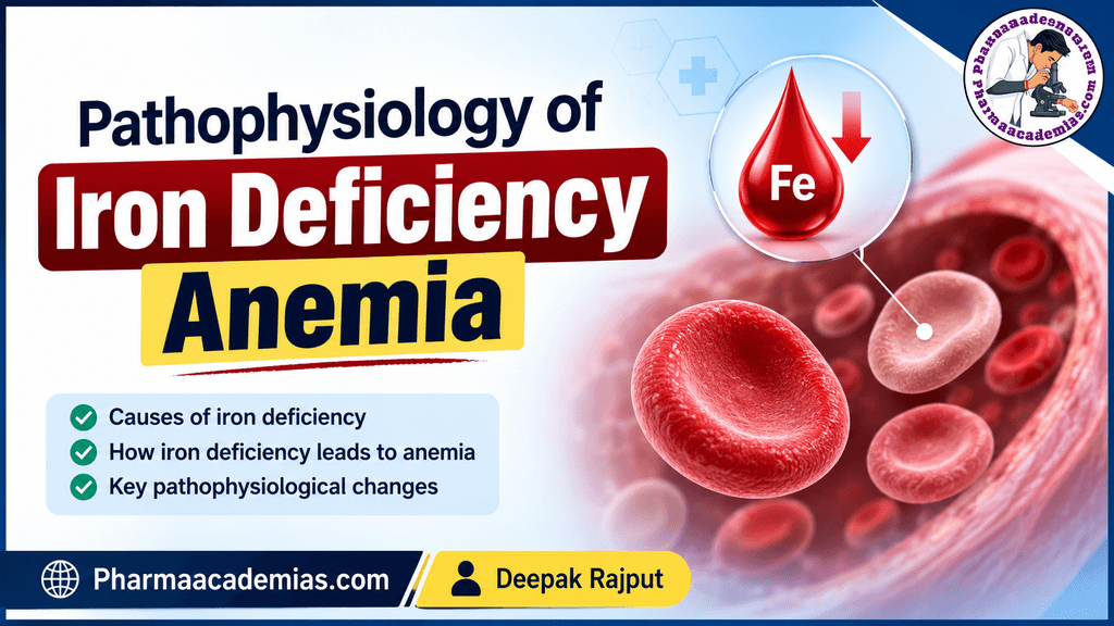
Iron is essential for the synthesis of hemoglobin, the protein responsible for transporting oxygen from the lungs to tissues. Therefore, iron deficiency primarily affects erythropoiesis, producing microcytic, hypochromic red blood cells and reduced hemoglobin concentration.
The pathophysiology of IDA can be broadly understood as a progression through:
Negative iron balance → depletion of iron stores → iron-restricted erythropoiesis → impaired hemoglobin synthesis → microcytic hypochromic anemia → tissue hypoxia and clinical manifestations.
2. Normal Iron Physiology
Understanding iron deficiency requires an understanding of normal iron metabolism.
2.1 Distribution of iron in the body
In a healthy adult, total body iron is approximately 3–4 g. It is distributed mainly among:
- Hemoglobin: approximately 60–70%
- Storage iron: ferritin and hemosiderin
- Myoglobin: approximately 10%
- Iron-containing enzymes: small amounts
- Plasma transferrin: very small circulating pool
The majority of functional iron is present in hemoglobin within circulating red blood cells.
2.2 Iron absorption
Dietary iron exists mainly in two forms:
- Heme iron
- Found primarily in meat and other animal products.
- Relatively efficiently absorbed.
- Non-heme iron
- Found predominantly in plant-based foods.
- Absorption is more strongly influenced by dietary and gastrointestinal factors.
Iron absorption primarily occurs in the duodenum and proximal jejunum.
Dietary iron is absorbed by intestinal enterocytes and subsequently exported into the circulation through ferroportin, the major cellular iron exporter.
2.3 Role of hepcidin
Hepcidin is the major hormone regulating systemic iron homeostasis.
It is produced primarily by the liver and binds to ferroportin, causing ferroportin internalization and degradation.
Consequently:
↑ Hepcidin → ↓ ferroportin activity → ↓ intestinal iron absorption → ↓ iron release from macrophages → ↓ plasma iron
Conversely:
↓ Hepcidin → ↑ ferroportin activity → ↑ iron absorption and iron release into circulation
In uncomplicated iron deficiency, hepcidin production generally decreases, allowing the intestine to increase iron absorption. However, if iron loss or insufficient intake continues, this compensatory mechanism cannot maintain adequate iron availability.
3. Basic Pathophysiology of Iron Deficiency Anemia
Iron deficiency develops when there is a persistent imbalance between:
Iron requirement + iron loss > iron absorption and availability
The major causes include:
- Chronic blood loss
- Inadequate dietary iron intake
- Increased physiological requirements
- Impaired gastrointestinal absorption
- Increased iron loss
- Certain chronic gastrointestinal disorders
The disease usually develops gradually rather than suddenly.
The progression can be divided into three major stages.
4. Stages of Iron Deficiency
Stage 1: Iron Depletion
The first stage is characterized by a reduction in the body’s iron stores.
Initially, the body uses stored iron from:
- Liver
- Spleen
- Bone marrow
- Macrophages
Stored iron is primarily present as:
- Ferritin
- Hemosiderin
During this stage, iron stores decline, but sufficient iron may still be available for hemoglobin synthesis.
Laboratory changes
The earliest significant laboratory abnormality is usually:
↓ Serum ferritin
Because ferritin reflects body iron stores, low ferritin is an important indicator of depleted iron reserves, although ferritin can be increased by inflammation because it is also an acute-phase reactant.
At this stage:
- Hemoglobin may remain normal.
- Hematocrit may remain normal.
- RBC morphology may remain relatively normal.
- Serum iron may still be within the reference range.
Therefore, the patient may have iron deficiency without anemia.
5. Stage 2: Iron-Restricted Erythropoiesis
As iron deficiency progresses, stored iron becomes insufficient to meet the requirements of developing erythroid cells in the bone marrow.
The bone marrow requires iron for the synthesis of hemoglobin.
When iron availability decreases:
↓ Iron availability → ↓ heme synthesis → ↓ hemoglobin synthesis
At this stage, erythrocyte production becomes increasingly iron restricted.
Important biochemical changes
Typically:
- ↓ Serum iron
- ↑ Total iron-binding capacity (TIBC)
- ↓ Transferrin saturation
- ↑ Transferrin receptor expression
- ↓ Ferritin
The bone marrow begins producing RBCs with progressively reduced hemoglobin content.
These cells may become:
- Smaller (microcytic)
- Paler (hypochromic)
However, significant anemia may not yet be present.
6. Stage 3: Iron Deficiency Anemia
If iron deficiency continues, hemoglobin synthesis becomes severely impaired.
The bone marrow cannot produce sufficient hemoglobin-containing RBCs.
Consequently:
Severe iron deficiency → impaired hemoglobin synthesis → reduced RBC hemoglobin content → microcytic hypochromic RBCs → decreased hemoglobin concentration → anemia
At this stage, hemoglobin and hematocrit become clearly reduced.
The blood contains RBCs that are:
Microcytic: RBCs are smaller than normal because inadequate hemoglobin synthesis results in reduced cellular maturation and division patterns.
Hypochromic: RBCs contain less hemoglobin, resulting in increased central pallor on peripheral blood smear.
7. Mechanism of Impaired Hemoglobin Synthesis
Hemoglobin consists of:
- Heme
- Globin chains
Iron is an essential component of the heme molecule.
The final step in heme biosynthesis involves incorporation of ferrous iron (Fe²⁺) into protoporphyrin IX to form heme.
This reaction is catalyzed by the enzyme:
Ferrochelatase
Therefore, inadequate intracellular iron results in reduced heme production.
The overall pathway can be simplified as:
Iron deficiency
↓
Reduced intracellular iron availability
↓
Reduced heme synthesis
↓
Reduced hemoglobin synthesis
↓
Reduced hemoglobin concentration per RBC
↓
Microcytic and hypochromic RBC production
8. Effect on Bone Marrow Erythropoiesis
The bone marrow continuously produces RBCs through the process of erythropoiesis.
Developing erythroid cells require:
- Iron
- Amino acids
- Vitamin B12
- Folate
- Erythropoietin
- Other micronutrients
Iron deficiency particularly affects hemoglobin production.
As iron availability falls, erythroid precursors cannot synthesize adequate hemoglobin.
This leads to:
Iron-restricted erythropoiesis → ineffective RBC production → reduced RBC hemoglobinization
The bone marrow may attempt to compensate by increasing erythropoietic activity through stimulation by erythropoietin (EPO).
However, increased erythropoiesis cannot adequately compensate when iron is unavailable.
9. Role of Tissue Hypoxia
Hemoglobin is responsible for oxygen transport.
When hemoglobin concentration decreases:
↓ Hemoglobin → ↓ oxygen-carrying capacity of blood → ↓ tissue oxygen delivery → tissue hypoxia
The body attempts to compensate for reduced oxygen delivery through several mechanisms.
9.1 Increased cardiac output
The cardiovascular system increases blood flow to tissues to compensate for reduced oxygen content.
This can produce:
- Palpitations
- Tachycardia
- Exercise intolerance
9.2 Redistribution of blood flow
Blood flow may preferentially support vital organs such as:
- Brain
- Heart
- Kidneys
while less essential tissues may receive relatively less oxygen.
9.3 Increased 2,3-BPG
Anemia can be associated with increased levels of 2,3-bisphosphoglycerate (2,3-BPG) in RBCs.
Increased 2,3-BPG decreases hemoglobin’s affinity for oxygen and facilitates oxygen release to peripheral tissues.
Thus:
Anemia → ↑ 2,3-BPG → rightward shift of oxygen dissociation curve → ↑ oxygen unloading
This is a compensatory mechanism.
10. Iron Deficiency and Cellular Function
Iron is not required only for hemoglobin.
It is also an essential component of several proteins and enzymes involved in:
- Mitochondrial energy production
- Electron transport
- Oxidative metabolism
- DNA synthesis
- Neurotransmitter metabolism
- Myoglobin function
Therefore, iron deficiency can produce symptoms even before severe anemia develops.
This explains why iron deficiency may be associated with:
- Fatigue
- Reduced concentration
- Cognitive difficulties
- Weakness
- Exercise intolerance
- Restless legs in some individuals
- Altered epithelial function
Thus, iron deficiency has biological consequences beyond the reduction in hemoglobin.
11. Gastrointestinal and Epithelial Changes
Iron deficiency can affect epithelial tissues.
One characteristic manifestation is atrophic changes in the oral and gastrointestinal mucosa.
Possible manifestations include:
- Glossitis
- Angular cheilitis
- Oral soreness
- Brittle nails
- Hair changes
- Dysphagia in some patients
A classic association is Plummer–Vinson syndrome, characterized by:
Iron deficiency anemia + dysphagia + esophageal webs
Although uncommon, it demonstrates the systemic effects of iron deficiency.
12. Pica and Iron Deficiency
Pica refers to the persistent craving for non-nutritive substances such as:
- Ice
- Soil
- Clay
- Chalk
- Paper
Pagophagia, the craving for ice, is particularly associated with iron deficiency.
The precise mechanism is not completely understood, but correction of iron deficiency often improves the behavior.
13. Pathophysiology of Common Causes
A. Chronic Blood Loss
Chronic blood loss is one of the most important causes of iron deficiency in adults.
Examples include:
- Gastrointestinal bleeding
- Heavy menstrual bleeding
- Recurrent epistaxis
- Parasitic infections
- Repeated blood donation
The mechanism is:
Chronic blood loss → continuous loss of iron-containing RBCs → depletion of iron stores → negative iron balance → iron deficiency → impaired erythropoiesis → anemia
In adults, unexplained iron deficiency anemia—particularly in men and postmenopausal women—requires evaluation for potential gastrointestinal blood loss.
14. Increased Iron Requirements
Iron requirements increase during periods of rapid growth and increased erythropoietic demand.
Important situations include:
Pregnancy
During pregnancy, iron requirements increase because iron is needed for:
- Expansion of maternal red cell mass
- Placental development
- Fetal growth
- Fetal iron stores
If dietary absorption and supplementation do not meet the increased requirement:
Increased demand → negative iron balance → depletion of iron stores → IDA
Infancy and adolescence
Rapid growth increases the demand for iron.
Adolescents may be particularly vulnerable because of:
- Rapid growth
- Increased blood volume
- Menstrual blood loss in menstruating individuals
15. Inadequate Iron Intake
Insufficient dietary iron intake can contribute to iron deficiency.
This may occur because of:
- Poor dietary diversity
- Malnutrition
- Low intake of iron-rich foods
- Certain restrictive diets without adequate iron planning
However, in many adults, significant IDA should not automatically be attributed to diet alone; ongoing blood loss or impaired absorption may also need consideration.
16. Malabsorption
Even when adequate iron is consumed, iron deficiency may occur if absorption is impaired.
Conditions associated with impaired iron absorption include:
- Celiac disease
- Certain gastric disorders
- Bariatric surgery
- Chronic gastrointestinal diseases
- Reduced gastric acidity in some circumstances
The mechanism is:
Impaired intestinal absorption → ↓ iron entering circulation → negative iron balance → depletion of stores → iron-restricted erythropoiesis → anemia
17. Iron Transport and Transferrin
After absorption, iron is transported through the blood primarily bound to transferrin.
Transferrin delivers iron to tissues, especially the bone marrow.
In iron deficiency:
↓ Body iron → ↑ transferrin production → ↑ TIBC
Because more transferrin is available but less iron is bound to it:
↓ Serum iron + ↑ TIBC → ↓ transferrin saturation
Transferrin saturation can therefore provide information about the amount of circulating iron available for erythropoiesis.
18. Ferritin and Storage Iron
Ferritin is the principal intracellular iron-storage protein and is also measured clinically as a marker of iron stores.
In uncomplicated iron deficiency:
↓ Iron stores → ↓ ferritin
However, ferritin is also an acute-phase reactant.
Inflammation can increase ferritin despite depleted or functionally unavailable iron stores.
Therefore, ferritin must sometimes be interpreted alongside:
- C-reactive protein (CRP)
- Serum iron
- Transferrin/TIBC
- Transferrin saturation
- Soluble transferrin receptor
- Reticulocyte hemoglobin, where available
Summary
Iron deficiency anemia develops through a progressive depletion of body iron stores followed by inadequate iron availability for hemoglobin synthesis.
The fundamental pathophysiological mechanism is:
Iron loss or inadequate iron supply → depletion of iron stores → reduced iron availability to bone marrow → impaired heme and hemoglobin synthesis → microcytic hypochromic RBC production → reduced hemoglobin concentration → decreased oxygen-carrying capacity → tissue hypoxia and compensatory physiological responses.
The disease progresses through three principal stages:
- Iron depletion – storage iron decreases, particularly ferritin.
- Iron-restricted erythropoiesis – insufficient iron reaches developing erythroid cells and hemoglobin synthesis becomes impaired.
- Iron deficiency anemia – hemoglobin falls and microcytic, hypochromic RBCs appear.
The clinical manifestations arise from both reduced oxygen delivery and the broader effects of iron deficiency on cellular enzymes, muscle function, epithelial tissues, and neurological processes.
Key concept to remember
The central defect in iron deficiency anemia is not simply a low hemoglobin level—it is inadequate iron availability for heme synthesis and erythropoiesis.
Editorial Note
This article has been carefully researched and written by Deepak Rajput with a focus on accuracy, clarity, and evidence-based healthcare information. Pharmaacademias.com