
Pathophysiology of Vertigo: Vertigo is a false sensation of movement, usually described as spinning, turning, tilting, or the surroundings moving when they are actually stationary. It is not a disease itself but a symptom caused by dysfunction of the vestibular system or its connections with the brain.
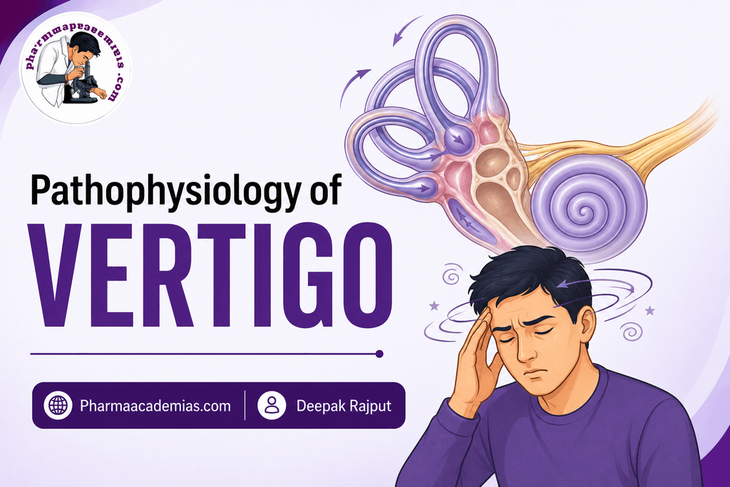
The vestibular system helps the body maintain balance, posture, spatial orientation, and stable vision during movement. It consists mainly of the inner-ear vestibular apparatus, vestibular nerve, brainstem, cerebellum, and their connections with the eyes and spinal cord.
Vertigo occurs when the brain receives conflicting or abnormal information about body movement and position, particularly from the vestibular system, vision, and proprioceptive system.
1. Normal Balance and Equilibrium
Maintenance of balance depends on the integration of information from three major sensory systems:
1. Vestibular system
Located in the inner ear, it detects:
- Head movement
- Angular acceleration
- Linear acceleration
- Gravity
- Head position
2. Visual system
The eyes provide information about:
- Position of the body in the environment
- Movement of the surroundings
- Orientation
3. Proprioceptive system
Receptors in:
- Muscles
- Tendons
- Joints
- Skin
provide information about the position and movement of different parts of the body.
The brain continuously compares information from these systems to maintain equilibrium.
Vestibular input + visual input + proprioceptive input → CNS integration → maintenance of balance
2. Vestibular Apparatus
The peripheral vestibular system is located in the inner ear and consists of:
Semicircular canals
There are three semicircular canals:
- Anterior/superior canal
- Posterior canal
- Horizontal/lateral canal
They detect angular or rotational acceleration of the head.
Otolith organs
The two otolith organs are:
- Utricle
- Saccule
They detect:
- Linear acceleration
- Head position relative to gravity
These structures contain specialized sensory hair cells.
3. Role of Hair Cells
Vestibular hair cells contain stereocilia and a kinocilium.
When the head moves, the fluid within the semicircular canals or the otolithic membrane moves relative to the sensory receptors.
This bends the hair cells and changes their electrical activity.
Head movement → movement of endolymph/otolithic structures → bending of hair cells → change in receptor potential → vestibular nerve signals
These signals are transmitted through the vestibular division of cranial nerve VIII (vestibulocochlear nerve) to the brainstem and cerebellum.
4. Basic Mechanism of Vertigo
Vertigo generally occurs when there is an asymmetry or mismatch in vestibular signaling.
For example, if the right and left vestibular organs normally send approximately equal signals:
Right vestibular activity ≈ Left vestibular activity → normal perception of head position
If one side suddenly becomes less active:
Right vestibular activity ↓
while:
Left vestibular activity remains normal
The brain interprets this difference as if the head is rotating.
Therefore:
Unequal vestibular signals → false perception of movement → vertigo
This is one of the major mechanisms responsible for acute peripheral vertigo.
5. Step-by-Step Pathophysiology of Vertigo
Step 1: Dysfunction of the Vestibular System
Vertigo may begin because of abnormalities in:
- Inner ear
- Vestibular nerve
- Brainstem
- Cerebellum
- Central vestibular pathways
Peripheral causes originate primarily from the inner ear or vestibular nerve, while central causes involve the brainstem, cerebellum, or higher CNS structures.
Step 2: Abnormal Vestibular Signals
Damage, inflammation, abnormal fluid pressure, displaced crystals, or other abnormalities can alter vestibular receptor activity.
This produces abnormal signals traveling through the vestibular nerve.
Vestibular dysfunction → abnormal afferent signals to CNS
Step 3: Sensory Mismatch
The brain normally compares information from:
- Vestibular system
- Eyes
- Proprioceptors
If these systems provide conflicting information, the CNS cannot accurately determine the body’s actual position and movement.
For example:
Vestibular system → “head is rotating”
while:
Eyes → “environment is stationary”
This sensory conflict can produce the sensation of spinning.
Step 4: Abnormal Vestibulo-Ocular Reflex
The vestibulo-ocular reflex (VOR) stabilizes vision during head movement by producing compensatory eye movements.
When vestibular signals become abnormal or asymmetric, inappropriate eye movements called nystagmus may occur.
Nystagmus consists of rhythmic, involuntary eye movements and is commonly associated with vestibular disorders.
Vestibular imbalance → abnormal VOR → nystagmus → visual instability
Step 5: Activation of Autonomic Centers
Vestibular pathways communicate with areas of the brainstem involved in autonomic functions.
Abnormal vestibular stimulation can therefore cause:
- Nausea
- Vomiting
- Sweating
- Pallor
- Increased heart awareness/palpitations in some individuals
Thus, vertigo is often accompanied by significant gastrointestinal and autonomic symptoms.
Step 6: Postural Instability
The vestibular system also communicates with the spinal cord through vestibulospinal pathways.
Abnormal vestibular activity can disturb:
- Muscle tone
- Postural reflexes
- Coordination
- Balance
This may cause:
- Unsteadiness
- Difficulty walking
- Falling or a tendency to fall
6. Pathophysiology in Peripheral Vertigo
Peripheral vertigo originates from the vestibular apparatus or vestibular nerve.
Common causes include:
- Benign paroxysmal positional vertigo (BPPV)
- Ménière’s disease
- Vestibular neuritis
- Labyrinthitis
- Other inner-ear disorders
Peripheral vertigo is often characterized by:
- Sudden spinning sensation
- Nausea and vomiting
- Nystagmus
- Imbalance
- Sometimes hearing loss or tinnitus, depending on the cause
7. Benign Paroxysmal Positional Vertigo (BPPV)
BPPV is one of the most common causes of peripheral vertigo.
Normally, calcium carbonate crystals called otoconia are located within the utricle.
Sometimes these crystals become displaced and enter a semicircular canal, most commonly the posterior canal.
Mechanism
Displaced otoconia → abnormal movement of endolymph during head movement → inappropriate stimulation of hair cells → abnormal vestibular signals → sensory mismatch → brief episodes of vertigo
The vertigo is typically triggered by changes in head position, such as:
- Turning in bed
- Looking upward
- Bending over
- Getting up from bed
8. Ménière’s Disease
Ménière’s disease is associated with endolymphatic hydrops, meaning abnormal accumulation of endolymph within the inner ear.
Increased endolymphatic pressure can disturb the normal function of vestibular and cochlear structures.
This may produce:
- Episodic vertigo
- Fluctuating hearing loss
- Tinnitus
- Ear fullness
A simplified sequence is:
Abnormal endolymph regulation → increased inner-ear fluid pressure → dysfunction of vestibular and cochlear structures → episodic vertigo ± hearing symptoms
9. Vestibular Neuritis
Vestibular neuritis involves dysfunction or inflammation affecting the vestibular nerve, often following a viral illness.
When vestibular function on one side is suddenly reduced:
Reduced vestibular activity on one side → vestibular asymmetry → brain interprets asymmetry as head movement → severe vertigo and nystagmus
Hearing is generally preserved because the cochlear portion of the eighth cranial nerve is not primarily affected.
10. Labyrinthitis
Labyrinthitis involves inflammation of the inner ear affecting vestibular structures and often the cochlear system.
Inflammation can disturb vestibular signaling and may also affect hearing.
Therefore, patients may develop:
- Vertigo
- Nausea and vomiting
- Imbalance
- Hearing loss
- Sometimes tinnitus
11. Central Vertigo
Central vertigo occurs when the structures responsible for processing vestibular information within the brainstem, cerebellum, or other CNS regions are affected.
Possible causes include:
- Stroke
- Multiple sclerosis
- Migraine-associated vestibular disorders
- Brain tumors
- Cerebellar disorders
- Other neurological conditions
In central vertigo, the problem is not primarily with the inner ear but with the processing and integration of vestibular information.
12. Peripheral vs Central Vertigo
| Feature | Peripheral Vertigo | Central Vertigo |
| Main site | Inner ear/vestibular nerve | Brainstem/cerebellum/CNS |
| Vertigo | Often intense | May be variable |
| Nystagmus | Often prominent | May have atypical patterns |
| Hearing symptoms | May occur | Usually absent, depending on cause |
| Nausea/vomiting | Often prominent | Variable |
| Postural imbalance | Usually present | May be severe |
| Neurological signs | Usually absent | May be present |
| Common examples | BPPV, Ménière’s disease, vestibular neuritis | Stroke, multiple sclerosis, cerebellar disorders |
Key Point
The central mechanism of vertigo is a mismatch or imbalance in sensory information about body movement and position. When vestibular signals from the two sides become unequal or are incorrectly processed, the brain may interpret the abnormal signals as actual head movement even when the person is stationary. This produces vertigo, while abnormal vestibulo-ocular and autonomic responses produce associated nystagmus, nausea, vomiting, and imbalance.
Editorial Note
This article has been carefully researched and written by Deepak Rajput with a focus on accuracy, clarity, and evidence-based healthcare information. Pharmaacademias.com