What is Dermatitis: Dermatitis is a broad term used to describe inflammation of the skin, resulting in symptoms such as redness, swelling, itching, pain, dryness, scaling, and sometimes blister formation. It is one of the most common dermatological conditions encountered in clinical practice and can affect individuals of all ages, from infants to the elderly. Although dermatitis is not usually life-threatening, it can significantly impair a person’s quality of life by causing persistent discomfort, disturbed sleep, emotional stress, and an increased risk of secondary skin infections. Pharmaacademias.com
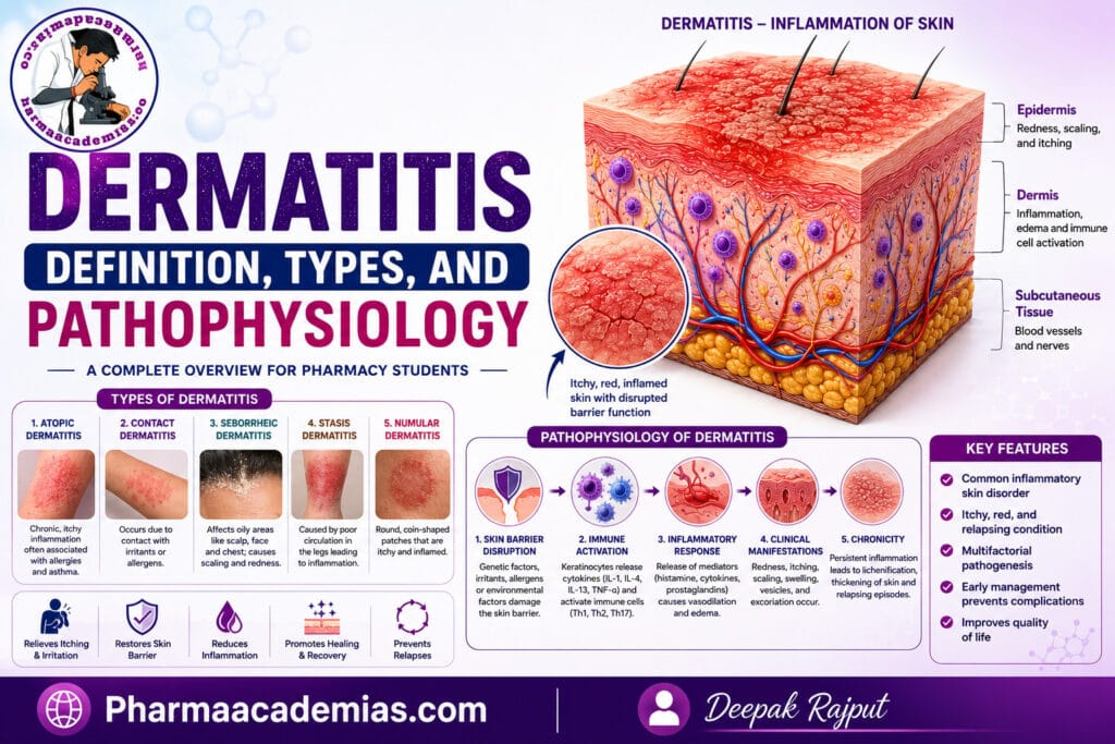
The skin normally functions as a protective barrier that prevents harmful microorganisms, allergens, chemicals, and excessive water loss. In dermatitis, this protective barrier becomes damaged or dysfunctional, allowing irritants, allergens, and microbes to penetrate the skin more easily. This activates the immune system, leading to inflammation and the characteristic symptoms of dermatitis.
Dermatitis is not a single disease but rather a group of inflammatory skin disorders with different causes and clinical presentations. Some forms are primarily caused by exposure to irritants or allergens, while others result from genetic predisposition, immune system dysfunction, or environmental factors. Regardless of the underlying cause, inflammation remains the central feature of all types of dermatitis.
What is Dermatitis
Dermatitis is defined as an inflammatory condition of the skin characterized by erythema (redness), itching, swelling, dryness, scaling, and, in some cases, vesicle or blister formation due to disruption of the skin barrier and activation of the immune system.
The condition may be acute, subacute, or chronic, depending on its duration and severity.
Types of Dermatitis
Several forms of dermatitis have been identified, each with distinct causes and clinical features.
1. Atopic Dermatitis (Atopic Eczema)
Atopic dermatitis is the most common chronic inflammatory skin disorder. It usually begins during infancy or early childhood but may also develop in adults.
It is strongly associated with genetic predisposition, immune dysregulation, and a defective skin barrier. Patients frequently have a personal or family history of asthma, allergic rhinitis, or food allergies, collectively known as the atopic triad.
The condition is characterized by intensely itchy, dry, inflamed skin that commonly affects the face in infants and the flexural areas (elbows, knees, neck) in older children and adults.
2. Contact Dermatitis
Contact dermatitis develops when the skin comes into direct contact with substances that either damage the skin or trigger an allergic reaction.
It is divided into two major forms.
Irritant Contact Dermatitis
This is the most common type and results from direct damage to the skin by irritants such as:
- Soaps and detergents
- Acids and alkalis
- Solvents
- Cleaning agents
- Frequent hand washing
The reaction does not require prior sensitization and may occur in any individual exposed to sufficient concentrations of the irritant.
Allergic Contact Dermatitis
This type results from a Type IV delayed hypersensitivity reaction mediated by T lymphocytes.
Common allergens include:
- Nickel jewelry
- Latex
- Cosmetics
- Perfumes
- Hair dyes
- Poison ivy
- Topical medications
Unlike irritant dermatitis, allergic contact dermatitis develops only after previous sensitization to the allergen.
3. Seborrheic Dermatitis
Seborrheic dermatitis primarily affects areas rich in sebaceous glands, including the scalp, face, eyebrows, ears, and chest.
The exact cause is unknown, but it is associated with:
- Overgrowth of Malassezia yeast
- Increased sebum production
- Immune response abnormalities
It presents with greasy yellow scales, redness, and itching.
In infants, it commonly appears as cradle cap.
4. Nummular Dermatitis
Nummular dermatitis is characterized by coin-shaped, itchy, inflamed plaques that commonly appear on the arms and legs.
The exact cause remains uncertain, although dry skin, trauma, and environmental factors may contribute.
5. Stasis Dermatitis
Stasis dermatitis develops due to chronic venous insufficiency, particularly in older adults.
Poor venous circulation causes fluid accumulation in the lower legs, resulting in inflammation, swelling, skin discoloration, and ulcer formation.
6. Dyshidrotic Dermatitis (Pompholyx)
This condition produces small, intensely itchy blisters on the palms, fingers, and soles of the feet.
Episodes are often triggered by stress, sweating, metal allergies, or seasonal changes.
7. Neurodermatitis (Lichen Simplex Chronicus)
Neurodermatitis develops due to repeated scratching or rubbing of the skin.
Continuous mechanical irritation causes thickened, leathery skin with exaggerated skin markings.
Pathophysiology of Dermatitis
The pathophysiology of dermatitis involves skin barrier dysfunction, immune system activation, inflammatory mediator release, and chronic inflammation. Although the exact mechanism varies among different types of dermatitis, several common pathological events occur.
1. Skin Barrier Dysfunction
The first and most important event in dermatitis is the breakdown of the skin’s protective barrier.
Under normal conditions, the epidermis—particularly the stratum corneum—acts as a waterproof shield that prevents excessive water loss while blocking the entry of microorganisms, allergens, and chemical irritants.
In dermatitis, this barrier becomes damaged due to:
- Genetic defects (especially mutations in the filaggrin gene)
- Mechanical injury
- Frequent washing
- Chemical exposure
- Environmental factors
Loss of barrier integrity leads to increased transepidermal water loss (TEWL), causing dry, cracked skin.
At the same time, allergens, irritants, and microbes gain easier access into the deeper layers of the skin.
2. Entry of Irritants and Allergens
Once the protective barrier is disrupted, foreign substances penetrate the epidermis.
These substances may include:
- Dust mites
- Pollens
- Animal dander
- Chemicals
- Metals
- Bacteria
- Fungi
- Viruses
Keratinocytes recognize these harmful agents and release inflammatory mediators.
3. Activation of Innate Immune Cells
Damaged keratinocytes release cytokines and signaling molecules such as:
- IL-1
- IL-6
- TNF-α
- TSLP (Thymic Stromal Lymphopoietin)
These cytokines activate:
- Dendritic cells
- Macrophages
- Mast cells
- Neutrophils
Activated dendritic cells capture allergens and migrate to nearby lymph nodes.
4. Activation of Adaptive Immune Response
In the lymph nodes, dendritic cells present antigens to naïve T lymphocytes.
Depending on the type of dermatitis, different T-helper cells become activated.
In Atopic Dermatitis
The immune response is predominantly Th2-mediated.
Th2 cells produce cytokines including:
- IL-4
- IL-5
- IL-13
- IL-31
These cytokines stimulate IgE production, eosinophil activation, and allergic inflammation.
In Allergic Contact Dermatitis
The immune response is mainly Th1 and Th17 mediated.
Sensitized T lymphocytes recognize the allergen and release inflammatory cytokines that recruit macrophages and other inflammatory cells.
Because this process requires previous sensitization, symptoms usually appear 24–72 hours after allergen exposure, representing a Type IV delayed hypersensitivity reaction.
5. Cytokine Release and Inflammation
Activated immune cells release numerous inflammatory cytokines.
Important cytokines include:
IL-4: Promotes IgE production by B lymphocytes and suppresses skin barrier protein synthesis.
IL-13: Acts together with IL-4 to impair epidermal barrier function and sustain allergic inflammation.
IL-31: IL-31 is strongly associated with itching (pruritus).
High IL-31 levels stimulate sensory nerves, producing severe itching that characterizes atopic dermatitis.
TNF-α: TNF-α amplifies inflammation by increasing vascular permeability and recruiting inflammatory cells.
IL-1 and IL-6: These cytokines promote fever, inflammation, and activation of additional immune cells.
6. Mast Cell Activation
Mast cells release:
- Histamine
- Leukotrienes
- Prostaglandins
Histamine causes:
- Intense itching
- Redness
- Vasodilation
- Increased vascular permeability
These changes explain why dermatitis lesions become swollen, warm, and itchy.
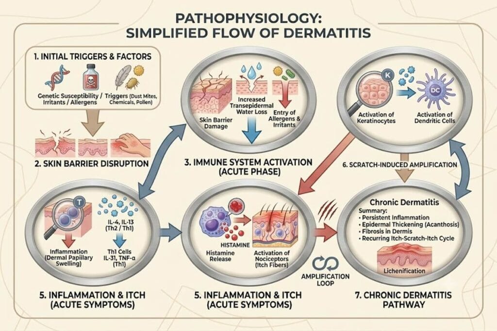
7. Chronic Itch–Scratch Cycle
One of the hallmark features of dermatitis is the itch–scratch cycle.
The sequence occurs as follows:
Inflammation stimulates sensory nerve endings, producing intense itching. The patient scratches the affected skin, causing additional skin damage. This further disrupts the skin barrier, allowing more allergens and irritants to enter. The immune response becomes stronger, leading to increased cytokine production and even more itching.
Over time, repeated scratching causes:
- Thickened skin (lichenification)
- Cracks
- Bleeding
- Secondary bacterial infection
This self-perpetuating cycle is responsible for chronic dermatitis.
8. Secondary Infection
The damaged skin barrier allows bacteria—especially Staphylococcus aureus—to colonize the skin.
These bacteria release toxins that further activate the immune system, increasing inflammation and worsening disease severity.
Secondary infections may produce:
- Pus formation
- Yellow crusting
- Increased redness
- Delayed healing
Simplified Flow of Dermatitis Pathophysiology
Clinical Features Resulting from the Pathophysiology
The underlying inflammatory mechanisms produce the characteristic symptoms of dermatitis, including:
- Redness (erythema)
- Intense itching (pruritus)
- Dry skin (xerosis)
- Swelling (edema)
- Scaling and flaking
- Vesicles or blisters in acute dermatitis
- Crusting and oozing
- Thickened skin (lichenification) in chronic dermatitis
- Cracks and fissures
- Increased susceptibility to bacterial infections
Complications of Dermatitis
If dermatitis is not adequately controlled, it may lead to several complications, such as:
- Chronic skin thickening (lichenification)
- Secondary bacterial infections (Staphylococcus aureus)
- Viral skin infections (e.g., eczema herpeticum)
- Fungal infections
- Sleep disturbances due to severe itching
- Anxiety, depression, and reduced quality of life
- Permanent skin discoloration (post-inflammatory hyperpigmentation or hypopigmentation)
Conclusion
Dermatitis is a common inflammatory skin disorder resulting from the interaction of skin barrier dysfunction, immune system activation, genetic susceptibility, and environmental triggers. The disease begins with disruption of the epidermal barrier, allowing irritants and allergens to penetrate the skin and activate both innate and adaptive immune responses. Cytokines such as IL-4, IL-13, IL-31, and TNF-α play central roles in maintaining inflammation and producing the hallmark symptom of intense itching, which leads to the characteristic itch–scratch cycle. This cycle perpetuates skin damage, chronic inflammation, and recurrent disease. Understanding the pathophysiology of dermatitis has led to the development of targeted therapies, including biologic agents that inhibit key inflammatory pathways, offering improved disease control and enhanced quality of life for affected patients.