Physiology of Muscle Contraction:: Muscle contraction is a physiological process in which skeletal muscle fibers generate force and shorten to produce movement. This process allows the body to perform voluntary activities such as walking, running, lifting objects, writing, and maintaining posture. Muscle contraction occurs through a highly coordinated interaction between the nervous system, muscle fibers, calcium ions, ATP (adenosine triphosphate), and the contractile proteins actin and myosin. The most widely accepted explanation for muscle contraction is the Sliding Filament Theory, proposed by Hugh Huxley and Andrew Huxley in 1954. Pharmaacademias.com
Skeletal muscles contract only when they receive signals from motor neurons. These nerve signals trigger a series of events known as excitation-contraction coupling, which converts an electrical impulse into a mechanical response. During contraction, the thin actin filaments slide over the thick myosin filaments, causing the sarcomere and ultimately the entire muscle to shorten. The muscle relaxes when the nerve stimulation stops, calcium ions return to storage, and the contractile proteins separate.
Steps in the Physiology of Muscle Contraction
Muscle contraction occurs in a sequence of well-coordinated events.
1. Generation of Nerve Impulse
The process begins when the brain or spinal cord sends a nerve impulse through a motor neuron to the skeletal muscle. This electrical signal, called an action potential, travels rapidly along the motor neuron until it reaches the neuromuscular junction, which is the point where the nerve communicates with the muscle fiber.
The action potential carries information from the nervous system and initiates muscle contraction only when required. Without this nerve signal, skeletal muscles remain relaxed.
2. Neuromuscular Junction and Release of Acetylcholine
The neuromuscular junction is the specialized connection between the motor neuron and the muscle fiber. When the action potential reaches the nerve terminal, voltage-gated calcium channels open, allowing calcium ions to enter the nerve ending.
The influx of calcium causes synaptic vesicles to release the neurotransmitter acetylcholine (ACh) into the synaptic cleft. Acetylcholine diffuses across the synaptic cleft and binds to receptors present on the sarcolemma (muscle cell membrane).
This binding opens sodium channels, allowing sodium ions to enter the muscle fiber. As a result, a new action potential is generated in the muscle cell membrane.
3. Propagation of Action Potential
The action potential spreads rapidly across the sarcolemma and then travels deep into the muscle fiber through narrow channels called transverse (T) tubules.
The T-tubules ensure that the electrical signal reaches every part of the muscle fiber almost simultaneously, allowing the entire muscle fiber to contract uniformly.
4. Release of Calcium Ions
The action potential travelling through the T-tubules stimulates the sarcoplasmic reticulum (SR), which is a specialized intracellular organelle that stores calcium ions.
In response, the sarcoplasmic reticulum releases a large amount of Ca²⁺ ions into the sarcoplasm. Calcium ions play a central role in initiating muscle contraction.
5. Binding of Calcium to Troponin
The thin actin filaments contain two important regulatory proteins:
- Troponin
- Tropomyosin
In a relaxed muscle, tropomyosin covers the active binding sites on actin, preventing myosin from attaching.
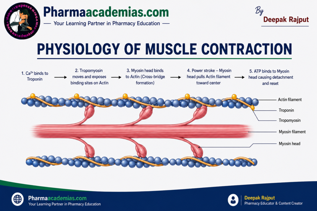
When calcium ions are released, they bind to troponin, causing a change in its shape. This conformational change moves tropomyosin away from the binding sites on actin, exposing the sites where myosin heads can attach.
6. Formation of Cross-Bridges
The exposed binding sites on actin allow energized myosin heads to attach firmly to actin filaments. This attachment forms structures known as cross-bridges.
ATP is required to energize the myosin heads before they bind to actin. The enzyme ATPase present in the myosin head hydrolyzes ATP into ADP and inorganic phosphate (Pi), storing energy that will be used during contraction.
7. Power Stroke
Once the cross-bridge is formed, the myosin head bends toward the center of the sarcomere, pulling the actin filament inward. This movement is known as the power stroke.
During the power stroke:
- ADP and Pi are released from the myosin head.
- Thin actin filaments slide toward the center of the sarcomere.
- The sarcomere becomes shorter.
- The muscle fiber contracts.
As millions of sarcomeres shorten simultaneously, the entire muscle shortens and generates force.
8. Detachment of Myosin Head
After the power stroke, a new ATP molecule binds to the myosin head.
The binding of ATP causes the myosin head to detach from actin. Without ATP, the myosin head remains attached to actin, leading to muscle stiffness, as seen in rigor mortis after death.
9. Reactivation of Myosin Head
The newly attached ATP is hydrolyzed by myosin ATPase into ADP and Pi.
This hydrolysis releases energy that re-cocks or reactivates the myosin head, preparing it for another cycle of attachment and power stroke.
As long as calcium ions remain available and ATP is continuously supplied, this cycle repeats many times each second.
10. Muscle Relaxation
Muscle relaxation begins when nerve impulses stop reaching the muscle.
Acetylcholine present in the synaptic cleft is rapidly broken down by the enzyme acetylcholinesterase, preventing further stimulation of the muscle fiber.
Calcium ions are actively pumped back into the sarcoplasmic reticulum by calcium pumps using ATP. As calcium concentration decreases, calcium dissociates from troponin, allowing tropomyosin to once again cover the active sites on actin.
Without exposed binding sites, cross-bridge formation stops, the muscle fibers return to their resting length, and the muscle relaxes.
Sliding Filament Theory
The sliding filament theory explains that muscle contraction occurs because actin filaments slide over myosin filaments, rather than because either filament shortens.
During contraction:
- Thin actin filaments move toward the center of the sarcomere.
- Thick myosin filaments remain in the same position.
- The sarcomere shortens.
- The I-band and H-zone become smaller.
- The A-band remains unchanged because the length of the myosin filament does not change.
Thus, muscle shortening results from increased overlap between actin and myosin filaments.
Role of ATP in Muscle Contraction
ATP is essential throughout the muscle contraction cycle. It provides the energy required for several important processes:
- Energizes the myosin heads before cross-bridge formation.
- Causes detachment of myosin from actin after the power stroke.
- Powers the calcium pumps that return calcium ions to the sarcoplasmic reticulum during relaxation.
- Maintains normal muscle metabolism and cellular activities.
Without ATP, muscles cannot contract or relax properly.
Role of Calcium in Muscle Contraction
Calcium ions act as the key regulator of muscle contraction.
Their main functions include:
- Binding to troponin.
- Moving tropomyosin away from actin.
- Exposing myosin-binding sites on actin.
- Initiating cross-bridge formation.
- Maintaining repeated contraction while calcium levels remain high.
When calcium is removed from the sarcoplasm, muscle contraction stops and relaxation begins.
Changes in the Sarcomere During Contraction
During muscle contraction, several structural changes occur within the sarcomere.
| Structure | Change During Contraction |
| Sarcomere | Shortens |
| Z-lines | Move closer together |
| I-band | Becomes narrower |
| H-zone | Becomes narrower or disappears |
| A-band | Remains unchanged |
| Actin filament | Does not shorten; slides inward |
| Myosin filament | Does not shorten |
Factors Required for Normal Muscle Contraction
Normal muscle contraction depends on several essential components:
- Healthy motor neurons.
- Functional neuromuscular junction.
- Adequate release of acetylcholine.
- Availability of calcium ions.
- Sufficient ATP production.
- Normal actin and myosin proteins.
- Proper electrolyte balance, especially sodium, potassium, and calcium.
Clinical Importance
Disorders affecting any step of muscle contraction can impair normal muscle function.
- Myasthenia gravis: An autoimmune disorder in which antibodies block acetylcholine receptors at the neuromuscular junction, leading to muscle weakness.
- Muscular dystrophy: A group of inherited disorders characterized by progressive degeneration and weakness of skeletal muscles.
- Tetany: Sustained muscle spasms caused by low blood calcium levels (hypocalcemia).
- Rigor mortis: Muscle stiffness that develops after death due to depletion of ATP, preventing myosin from detaching from actin.
Conclusion
Muscle contraction is a complex but highly coordinated physiological process that enables voluntary movement. It begins with a nerve impulse, followed by the release of acetylcholine, generation of an action potential, release of calcium ions, and interaction between actin and myosin through the sliding filament mechanism. ATP provides the energy required for every stage of contraction and relaxation, while calcium acts as the key regulator that initiates the process. The coordinated action of the nervous system, contractile proteins, calcium ions, and ATP allows skeletal muscles to produce force, maintain posture, stabilize joints, and perform all voluntary body movements.
Editorial Note
This article has been carefully researched and written by Deepak Rajput with a focus on accuracy, clarity, and evidence-based healthcare information.