
Organization of Skeletal Muscle: Skeletal muscle is a specialized type of muscle tissue that is attached to bones and is responsible for voluntary body movements. It is called skeletal muscle because it is connected to the skeleton through tendons and works under conscious control. Skeletal muscles make up about 40–50% of the total body weight in a healthy adult. They are striated (striped in appearance), multinucleated, and capable of rapid and forceful contraction. Besides producing movement, skeletal muscles help maintain posture, stabilize joints, generate body heat, and support breathing.
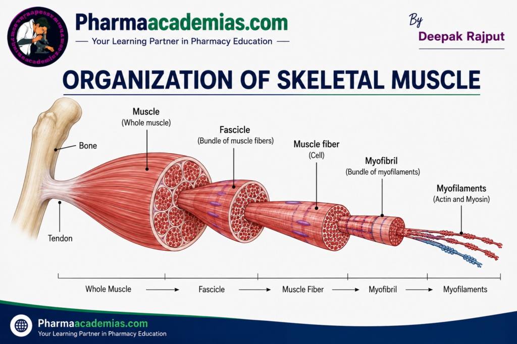
The organization of skeletal muscle is highly structured. Each muscle is made up of several layers, beginning with the whole muscle and ending with tiny protein filaments responsible for muscle contraction. This hierarchical organization allows muscles to contract efficiently and generate force.
Levels of Organization of Skeletal Muscle
The skeletal muscle is organized into the following levels:
Whole Muscle → Fascicle → Muscle Fiber (Muscle Cell) → Myofibril → Sarcomere → Myofilaments (Actin and Myosin)
Each level has a specific structure and function that contributes to muscle contraction.
1. Whole Muscle
The whole skeletal muscle is the organ that is visible to the naked eye. It is composed of many bundles of muscle fibers along with connective tissues, blood vessels, and nerves. Each muscle is attached to bones by strong fibrous structures called tendons, which transmit the force generated during muscle contraction to produce movement.
The entire muscle is covered by a dense connective tissue layer called the epimysium. The epimysium protects the muscle, maintains its shape, and separates it from surrounding tissues. Blood vessels and nerves enter the muscle through this layer to supply nutrients, oxygen, and nerve signals.
Examples of whole skeletal muscles include the biceps brachii, triceps brachii, quadriceps femoris, gastrocnemius, and deltoid muscles.
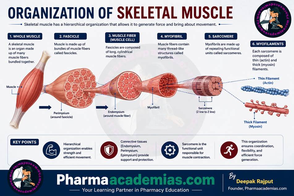
2. Fascicles
Inside the whole muscle, the muscle fibers are arranged into bundles known as fascicles. Each fascicle contains several muscle fibers grouped together.
Every fascicle is surrounded by another connective tissue layer called the perimysium. The perimysium protects the fascicles, supports blood vessels and nerves, and helps distribute the force generated during muscle contraction throughout the muscle.
The arrangement of fascicles determines the strength and range of movement of different muscles. Depending on their arrangement, muscles may be parallel, fusiform, circular, convergent, or pennate.
3. Muscle Fiber (Muscle Cell)
A muscle fiber is a single elongated muscle cell. Unlike most body cells, skeletal muscle fibers are very long and contain multiple nuclei located near the cell membrane. Some muscle fibers may extend the entire length of a muscle.
Each muscle fiber is enclosed by a thin connective tissue layer called the endomysium, which surrounds individual muscle cells and provides structural support. The muscle cell membrane is called the sarcolemma, while the cytoplasm is known as the sarcoplasm.
The sarcoplasm contains glycogen, mitochondria, myoglobin, enzymes, and numerous myofibrils. Myoglobin stores oxygen needed during muscle activity, while mitochondria produce ATP, the energy required for muscle contraction.
4. Myofibrils
Inside each muscle fiber are hundreds to thousands of long cylindrical structures called myofibrils. They are the contractile elements of the muscle cell and occupy most of the cell’s interior.
Myofibrils are arranged parallel to each other and extend the entire length of the muscle fiber. Their orderly arrangement produces the characteristic light and dark bands seen under the microscope, giving skeletal muscle its striated appearance.
Each myofibril consists of repeating contractile units called sarcomeres.
5. Sarcomere
The sarcomere is the smallest structural and functional unit of skeletal muscle contraction. It extends from one Z-line (Z-disc) to the next Z-line. During muscle contraction, the sarcomere shortens, leading to shortening of the entire muscle.
A sarcomere contains overlapping thick and thin filaments arranged in a highly organized pattern.
Its important parts include:
- Z-line (Z-disc): Forms the boundary of each sarcomere and anchors the thin filaments.
- I-band: Contains only thin (actin) filaments and appears lighter under the microscope.
- A-band: Contains the entire length of thick (myosin) filaments and appears darker.
- H-zone: Central region containing only thick filaments.
- M-line: Located in the center of the H-zone and helps stabilize the thick filaments.
According to the sliding filament theory, muscle contraction occurs when thin actin filaments slide over thick myosin filaments, shortening the sarcomere without changing the length of the filaments themselves.
6. Myofilaments
The myofilaments are the protein filaments that directly produce muscle contraction. There are two main types:
Thick Filaments (Myosin): Thick filaments are composed mainly of the protein myosin. Each myosin molecule has a long tail and two globular heads. The heads contain ATPase enzyme activity, which breaks down ATP to provide energy for contraction. During contraction, the myosin heads bind to actin and pull the thin filaments toward the center of the sarcomere.
Thin Filaments (Actin): Thin filaments are mainly composed of actin, along with the regulatory proteins troponin and tropomyosin. Actin provides binding sites for myosin heads. Tropomyosin blocks these binding sites when the muscle is relaxed, while troponin binds calcium ions and moves tropomyosin away during contraction, allowing actin and myosin to interact.
Connective Tissue Coverings of Skeletal Muscle
Three connective tissue layers surround skeletal muscle and provide support, protection, and organization.
Epimysium:The epimysium surrounds the entire muscle. It protects the muscle, maintains its shape, and blends with tendons that attach the muscle to bones.
Perimysium: The perimysium surrounds each fascicle. It carries blood vessels and nerves into the muscle and helps distribute the force of contraction.
Endomysium: The endomysium surrounds each individual muscle fiber. It provides support to muscle cells and contains fine capillaries and nerve endings that supply the muscle fiber.
Blood Supply and Nerve Supply
Skeletal muscles have a rich blood supply because they require a continuous supply of oxygen and nutrients to produce energy. Arteries branch into capillaries that surround individual muscle fibers, while veins remove carbon dioxide and metabolic waste products.
Each skeletal muscle is supplied by motor neurons, which stimulate muscle contraction. The point where the motor neuron communicates with the muscle fiber is called the neuromuscular junction. Here, the neurotransmitter acetylcholine (ACh) is released, initiating muscle contraction.
Functions of Skeletal Muscle
The organized structure of skeletal muscle enables it to perform several important functions:
- Produces voluntary movements of the body.
- Maintains posture and body position.
- Stabilizes joints during movement.
- Generates heat to help maintain body temperature.
- Supports breathing through muscles such as the diaphragm and intercostal muscles.
- Protects internal organs by forming muscular walls around them.
- Stores glycogen as an energy reserve for muscle activity.
Summary of Skeletal Muscle Organization
| Level of Organization | Description | Main Function |
| Whole Muscle | Entire muscle organ covered by epimysium | Produces movement and generates force |
| Fascicle | Bundle of muscle fibers surrounded by perimysium | Organizes muscle fibers and distributes force |
| Muscle Fiber | Individual muscle cell surrounded by endomysium | Contracts in response to nerve stimulation |
| Myofibril | Rod-like contractile structure inside the muscle fiber | Contains sarcomeres responsible for contraction |
| Sarcomere | Basic contractile unit between two Z-lines | Shortens during muscle contraction |
| Myofilaments | Actin (thin) and myosin (thick) protein filaments | Directly generate muscle contraction through the sliding filament mechanism |
Conclusion
The organization of skeletal muscle follows a highly ordered hierarchy, from the whole muscle down to microscopic protein filaments. Each structural level plays a vital role in ensuring efficient muscle contraction, force generation, and movement. The connective tissue coverings, rich blood supply, and nerve supply work together to maintain muscle health and function. This well-organized arrangement enables skeletal muscles to perform essential activities such as movement, posture maintenance, breathing, joint stabilization, and heat production, making them indispensable for normal human life.
Editorial Note
This article has been carefully researched and written by Deepak Rajput with a focus on accuracy, clarity, and evidence-based healthcare information.