Salient Features and Functions of Bones: The human skeletal system is divided into two major parts: the axial skeletal system and the appendicular skeletal system. Together, these two divisions form the body’s framework, providing support, protection, movement, and stability. Although both systems are made of bones, they differ in their location, structure, and functions. The axial skeleton mainly forms the central axis of the body and protects vital organs, while the appendicular skeleton is responsible for body movements and locomotion. Pharmaacademias.com
Axial Skeletal System
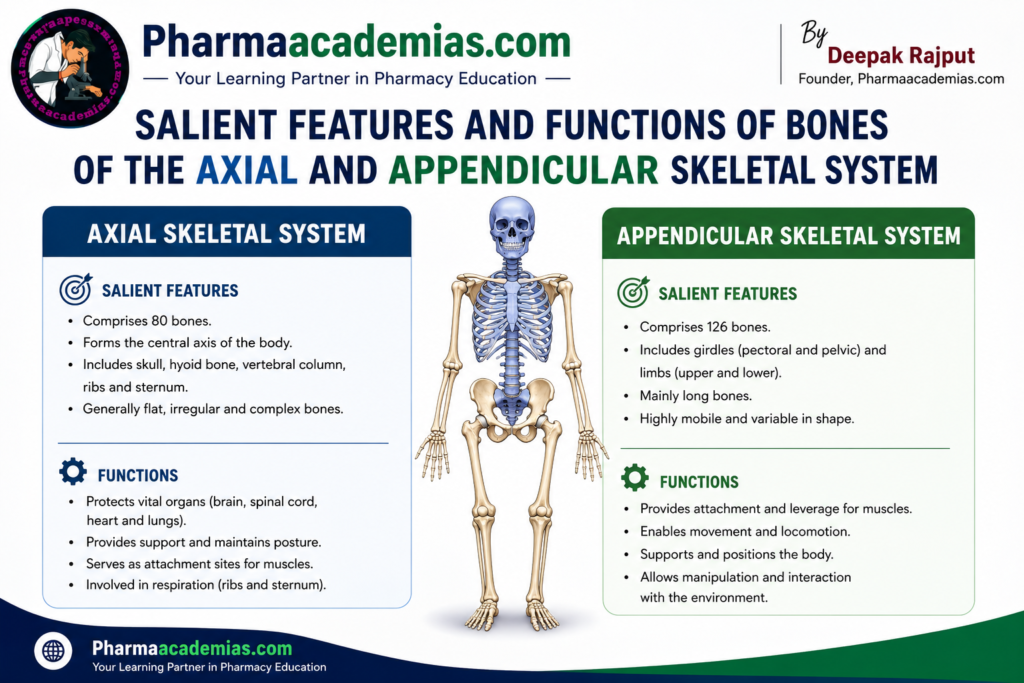
The axial skeleton forms the central supporting axis of the body. It consists of 80 bones, including the skull, hyoid bone, auditory ossicles, vertebral column, sternum, and ribs. These bones are arranged to protect the brain, spinal cord, heart, lungs, and other vital organs while providing attachment sites for muscles involved in posture, breathing, and head movement.
Salient Features of the Axial Skeleton
The axial skeleton is located along the body’s midline and forms the central framework. It contains 80 bones and is primarily designed for strength and protection rather than extensive movement. The bones are interconnected in a way that provides stability while allowing limited flexibility. Most bones of the axial skeleton are firmly connected to each other, making this division highly resistant to injury.
The axial skeleton serves as the point of attachment for many muscles that control the movements of the head, neck, chest, and back. It also provides support for maintaining an upright posture and protecting delicate organs that are essential for life.
The major components of the axial skeleton include:
- Skull (22 bones)
- Hyoid bone (1 bone)
- Auditory ossicles (6 bones)
- Vertebral column (26 bones in adults)
- Sternum (1 bone)
- Twelve pairs of ribs (24 bones)
Functions of the Bones of the Axial Skeleton
Skull
The skull is one of the most important structures of the axial skeleton. It surrounds and protects the brain from injury. The facial bones provide the basic framework of the face, support the eyes, nose, and mouth, and give attachment to muscles responsible for chewing, speaking, swallowing, and facial expressions. The skull also houses the organs of vision, hearing, smell, and taste.
Hyoid Bone
The hyoid bone supports the tongue and provides attachment for muscles involved in swallowing and speech. Unlike other bones, it does not directly articulate with another bone, making it unique within the skeletal system.
Auditory Ossicles
The auditory ossicles are the smallest bones in the human body. They transmit sound vibrations from the eardrum to the inner ear, allowing hearing. These tiny bones increase the efficiency of sound transmission and are essential for normal hearing.
Vertebral Column
The vertebral column supports the weight of the head and trunk while protecting the spinal cord. It provides flexibility that allows bending, twisting, and rotation of the body. The vertebrae also serve as attachment sites for muscles and ligaments that maintain posture and body balance.
Sternum
The sternum forms the front part of the thoracic cage. It provides attachment for ribs and clavicles and protects important organs such as the heart, major blood vessels, and lungs.
Ribs
The ribs form the protective cage around the chest. They protect the heart and lungs from injury while also participating in the breathing process by expanding and contracting during respiration.
Appendicular Skeletal System
The appendicular skeleton consists of 126 bones and includes the bones of the upper limbs, lower limbs, shoulder girdle, and pelvic girdle. It is primarily responsible for movement, balance, and locomotion. Compared to the axial skeleton, the appendicular skeleton contains more movable joints, allowing greater flexibility and a wider range of motion.
Salient Features of the Appendicular Skeleton
The appendicular skeleton includes all the bones that form the limbs and the girdles connecting them to the axial skeleton. It contains 126 bones, making it the larger division of the skeletal system.
The appendicular skeleton is specially adapted for movement. The bones are connected through highly movable joints that allow activities such as walking, running, climbing, lifting, throwing, and grasping objects. The shoulder girdle provides exceptional mobility to the upper limbs, whereas the pelvic girdle provides strength and stability for weight-bearing and locomotion.
The appendicular skeleton is composed of:
- Pectoral (shoulder) girdles – 4 bones
- Upper limbs – 60 bones
- Pelvic girdle – 2 hip bones
- Lower limbs – 60 bones
Functions of the Bones of the Appendicular Skeleton
Pectoral (Shoulder) Girdle
The pectoral girdle connects the upper limbs to the axial skeleton. It provides flexibility and a wide range of movement while serving as an attachment site for muscles of the shoulder, chest, and upper back.
Humerus
The humerus forms the upper arm and acts as a lever for muscles, allowing movements at both the shoulder and elbow joints. It is essential for lifting, pushing, pulling, and rotating the arm.
Radius and Ulna
These two bones form the forearm. They allow movements such as bending and straightening of the elbow as well as rotation of the forearm during actions like turning a doorknob or using a screwdriver.
Carpals, Metacarpals, and Phalanges
These bones form the wrist, hand, and fingers. They provide flexibility, grip strength, and fine motor control necessary for writing, typing, drawing, eating, and handling delicate objects.
Pelvic Girdle
The pelvic girdle connects the lower limbs to the axial skeleton. It supports the weight of the upper body, protects pelvic organs such as the urinary bladder and reproductive organs, and provides attachment for strong muscles involved in walking and standing.
Femur
The femur is the longest and strongest bone in the human body. It supports body weight and plays a major role in standing, walking, running, and jumping.
Patella
The patella protects the knee joint and improves the efficiency of the thigh muscles by increasing the leverage of the quadriceps muscle during leg extension.
Tibia and Fibula
The tibia is the main weight-bearing bone of the lower leg, while the fibula provides muscle attachment and contributes to ankle stability. Together they enable standing, walking, and maintaining balance.
Tarsals, Metatarsals, and Phalanges
These bones form the ankle, foot, and toes. They support body weight, absorb shock, maintain balance, and help in walking, running, and jumping.
Comparison Between Axial and Appendicular Skeleton
| Feature | Axial Skeleton | Appendicular Skeleton |
| Number of Bones | 80 | 126 |
| Position | Central axis of the body | Limbs and girdles |
| Main Function | Support and protection | Movement and locomotion |
| Major Components | Skull, vertebral column, ribs, sternum | Shoulder girdle, upper limbs, pelvic girdle, lower limbs |
| Type of Movement | Limited movement | Wide range of movement |
| Primary Role | Protects vital organs | Enables mobility and manipulation |
Overall Functions of the Skeletal System
The bones of both the axial and appendicular skeleton work together to perform several essential functions:
- Provide shape and structural support to the body.
- Protect vital organs such as the brain, spinal cord, heart, lungs, and pelvic organs.
- Enable movement by acting as levers for skeletal muscles.
- Maintain body posture and balance.
- Store essential minerals, especially calcium and phosphorus.
- Produce red blood cells, white blood cells, and platelets in the red bone marrow.
- Store fat in the yellow bone marrow as an energy reserve.
- Provide attachment sites for muscles, ligaments, and tendons.
- Absorb mechanical stress and distribute body weight during movement.
Conclusion
The axial skeleton and appendicular skeleton are two complementary divisions of the human skeletal system. The axial skeleton provides a strong central framework that supports the body and protects vital organs such as the brain, spinal cord, heart, and lungs. In contrast, the appendicular skeleton is specialized for movement, allowing the body to perform a wide variety of activities ranging from simple daily tasks to complex physical actions. Together, these two divisions ensure that the human body remains stable, protected, flexible, and capable of efficient movement while also contributing to mineral storage and blood cell production.
Editorial Note
This article has been carefully researched and written by Deepak Rajput with a focus on accuracy, clarity, and evidence-based healthcare information.