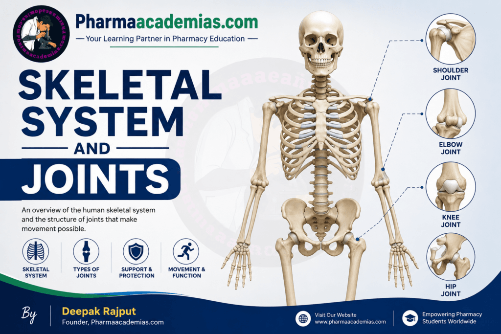
Skeletal System and Joints: The skeletal system is the framework of the human body. It is made up of bones, cartilage, ligaments, and joints that work together to support the body, protect internal organs, and help in movement. An adult human has 206 bones, while a newborn baby has around 270 bones. As a child grows, some bones fuse together, reducing the total number to 206. Without the skeletal system, the human body would not be able to maintain its shape or perform movements. It also plays an important role in storing minerals such as calcium and phosphorus and producing blood cells inside the bone marrow. Pharmaacademias.com
Bones are living tissues that are constantly changing. They are supplied with blood vessels and nerves, which help them grow, repair, and maintain their strength. The skeletal system works closely with muscles, tendons, and joints to produce movement. When muscles contract, they pull on bones through tendons, and the joints allow the bones to move smoothly. This coordination enables us to perform everyday activities such as walking, running, lifting, writing, and even smiling.
Joints are the places where two or more bones meet. They allow different types of movements depending on their structure and location. Some joints provide a wide range of motion, while others are almost immovable. Cartilage covers the ends of bones at joints, reducing friction and preventing damage during movement. Ligaments connect bones to other bones and provide stability to the joints. Synovial fluid, present in many joints, acts as a lubricant and helps the bones move freely without excessive wear.
Divisions of the Skeletal System
The human skeletal system is divided into two major parts:
Both divisions have different functions, but together they form a complete framework for the body.
1. Axial Skeleton
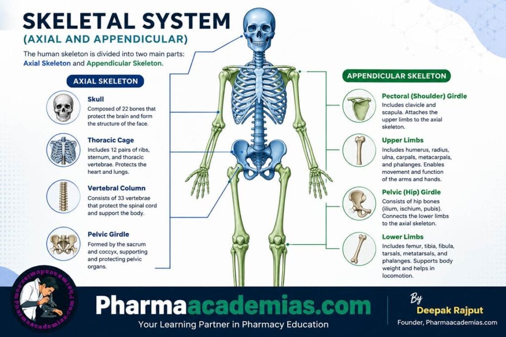
The axial skeleton forms the central axis of the body. It consists of 80 bones and mainly provides support and protection for the brain, spinal cord, and organs located in the chest.
The axial skeleton acts as the body’s main supporting structure. It helps maintain posture, protects delicate organs, and provides attachment sites for muscles involved in breathing, head movement, and maintaining body balance. Since it forms the central framework of the body, all other parts of the skeleton are connected to it.
The axial skeleton includes the following parts:
Skull
The skull is made up of 22 bones that protect the brain and form the structure of the face. The bones of the skull are joined together by immovable joints called sutures, making the skull very strong. The skull protects the brain from injury and also supports the eyes, nose, ears, and mouth. The facial bones provide attachment for muscles responsible for chewing, speaking, and facial expressions.
Hyoid Bone
The hyoid bone is a small, U-shaped bone located in the neck. It is unique because it does not directly articulate with any other bone. Instead, it is suspended by muscles and ligaments. The hyoid bone supports the tongue and helps in swallowing and speech.
Auditory Ossicles
The auditory ossicles are the smallest bones in the human body and are located in the middle ear. There are three bones in each ear: the malleus, incus, and stapes. These tiny bones transmit sound vibrations from the eardrum to the inner ear, making hearing possible.
Vertebral Column
The vertebral column, commonly known as the spine or backbone, consists of 33 vertebrae, although in adults some of these bones fuse together. The vertebral column supports the body’s weight, maintains posture, and protects the spinal cord. It also allows flexibility, enabling movements such as bending, twisting, and turning.
The vertebral column is divided into five regions:
- Cervical vertebrae (7)
- Thoracic vertebrae (12)
- Lumbar vertebrae (5)
- Sacrum (5 fused vertebrae)
- Coccyx (4 fused vertebrae)
Thoracic Cage
The thoracic cage is made up of the sternum (breastbone) and 12 pairs of ribs. It protects vital organs such as the heart and lungs. The rib cage also assists in the breathing process by expanding and contracting during inhalation and exhalation.
2. Appendicular Skeleton
The appendicular skeleton consists of 126 bones and includes the bones of the upper limbs, lower limbs, and the girdles that attach these limbs to the axial skeleton. Its primary function is to facilitate movement and provide support for activities such as walking, running, lifting, climbing, and grasping objects.
Unlike the axial skeleton, which mainly provides protection, the appendicular skeleton is specially designed for mobility. Most of the body’s joints that allow a wide range of movements are located in this division.
The appendicular skeleton includes the following parts:
Pectoral (Shoulder) Girdle
The pectoral girdle connects the upper limbs to the axial skeleton. It consists of two clavicles (collarbones) and two scapulae (shoulder blades). The shoulder girdle provides flexibility and allows a wide range of arm movements.
Upper Limbs
Each upper limb contains 30 bones, including:
- Humerus
- Radius
- Ulna
- Carpals (wrist bones)
- Metacarpals (hand bones)
- Phalanges (finger bones)
These bones enable activities requiring precision, such as writing, typing, lifting, throwing, and holding objects.
Pelvic Girdle
The pelvic girdle is formed by two hip bones that connect the lower limbs to the vertebral column. It supports the body’s weight, protects pelvic organs such as the urinary bladder and reproductive organs, and provides attachment for powerful muscles involved in walking and standing.
Lower Limbs
Each lower limb also contains 30 bones, including:
- Femur
- Patella
- Tibia
- Fibula
- Tarsals (ankle bones)
- Metatarsals (foot bones)
- Phalanges (toe bones)
The bones of the lower limbs are larger and stronger than those of the upper limbs because they support the body’s weight and provide stability during standing, walking, running, and jumping.
Joints
A joint, also known as an articulation, is the point where two or more bones meet. Joints allow movement and provide flexibility while maintaining the stability of the skeleton. Different joints permit different ranges of movement depending on their structure and function.
Joints are supported by ligaments, cartilage, muscles, tendons, and synovial fluid, all of which help reduce friction, absorb shock, and maintain smooth movement.
Joints are classified into three main types:
Fibrous Joints
Fibrous joints are connected by dense connective tissue and allow little or no movement. They provide stability and strength. The sutures of the skull are examples of fibrous joints.
Cartilaginous Joints
Cartilaginous joints are connected by cartilage and allow limited movement. They provide flexibility while maintaining stability. Examples include the joints between vertebrae and the pubic symphysis.
Synovial Joints
Synovial joints are the most common and freely movable joints in the human body. They contain a joint cavity filled with synovial fluid that lubricates the joint and reduces friction. Articular cartilage covers the ends of bones, allowing smooth movement.
Examples of synovial joints include:
- Ball-and-socket joint (shoulder and hip)
- Hinge joint (elbow and knee)
- Pivot joint (neck)
- Saddle joint (thumb)
- Condyloid joint (wrist)
- Gliding joint (between carpal bones)
Summary
The skeletal system provides the body with shape, support, and protection while also enabling movement. It consists of bones, joints, cartilage, and ligaments that function together as an integrated system. The skeleton is divided into the axial skeleton, which forms the body’s central framework and protects vital organs, and the appendicular skeleton, which includes the limbs and girdles responsible for movement. Joints connect bones and make body movements possible by allowing varying degrees of flexibility and stability. Together, the skeletal system and joints form the foundation of the musculoskeletal system, enabling the human body to perform both simple daily activities and complex physical movements.