Mechanism of Inflammation: Inflammation is a protective biological response of living, vascularized tissues to harmful stimuli such as infection, tissue injury, trauma, chemical irritation, toxins, and foreign bodies. Its primary purpose is to eliminate the initial cause of injury, remove damaged cells and tissues, and initiate tissue repair.
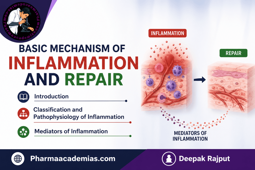
Inflammation is not itself a disease. It is an important component of the body’s defense system. However, when inflammation is excessive, prolonged, or inappropriately activated, it can cause tissue damage and contribute to diseases such as rheumatoid arthritis, atherosclerosis, asthma, inflammatory bowel disease, and cancer.
The classical features of inflammation are:
- Rubor – redness
- Calor – heat
- Tumor – swelling
- Dolor – pain
- Functio laesa – loss of function
These manifestations result primarily from changes in blood flow, vascular permeability, leukocyte recruitment, and release of inflammatory mediators.
2. Objectives of Inflammation
Inflammation performs several important functions:
- Recognition of injury or infection
- Removal of the harmful stimulus
- Elimination of microorganisms
- Removal of dead and damaged cells
- Prevention of further tissue damage
- Initiation of tissue repair
- Restoration of normal tissue structure and function
3. Causes of Inflammation
Inflammation may be initiated by several types of stimuli.
3.1 Infectious agents
These include:
- Bacteria
- Viruses
- Fungi
- Parasites
- Protozoa
Microbial products can activate immune cells and initiate inflammatory responses.
3.2 Physical agents
Examples include:
- Heat
- Cold
- Radiation
- Mechanical trauma
- Burns
- Electrical injury
3.3 Chemical agents
Examples include:
- Acids
- Alkalis
- Toxic chemicals
- Irritant substances
- Environmental pollutants
3.4 Tissue necrosis
Dead or damaged cells release intracellular molecules that activate inflammatory responses.
3.5 Foreign bodies
Examples include:
- Splinters
- Dust
- Sutures
- Crystals
- Particulate materials
3.6 Immune reactions
Inflammation can result from abnormal immune responses, including:
- Autoimmune diseases
- Allergic reactions
- Hypersensitivity reactions
4. Classification of Inflammation
Inflammation is primarily classified into:
- Acute inflammation
- Chronic inflammation
A simplified comparison is given below.
| Feature | Acute inflammation | Chronic inflammation |
| Onset | Rapid | Slow/insidious |
| Duration | Minutes to days | Weeks to years |
| Major cells | Neutrophils | Macrophages, lymphocytes, plasma cells |
| Vascular changes | Prominent | Usually less prominent |
| Exudation | Prominent | Variable |
| Tissue injury | Usually limited | Often progressive |
| Repair | Usually begins rapidly | Fibrosis/remodeling common |
| Typical examples | Acute appendicitis, cellulitis | Tuberculosis, rheumatoid arthritis |
5. Acute Inflammation
Acute inflammation is the early and relatively short-lived response to tissue injury or infection.
It is characterized by:
- Vasodilation
- Increased blood flow
- Increased vascular permeability
- Exudation of plasma
- Migration of leukocytes, particularly neutrophils
- Activation of inflammatory mediators
5.1 Vascular Changes
The vascular response occurs in several stages.
A. Transient vasoconstriction: Immediately after injury, there may be a very brief period of vasoconstriction.This is usually followed rapidly by vasodilation.
B. Vasodilation
Arterioles dilate, resulting in:
- Increased blood flow
- Increased tissue temperature
- Redness
This contributes to:
Rubor + Calor
Histamine, nitric oxide, and other mediators contribute to vasodilation.
C. Increased vascular permeability: The endothelial cells lining blood vessels become more permeable.Fluid and plasma proteins move from the blood into the surrounding tissue.
This produces:
Edema → Tumor
The protein-rich inflammatory fluid is called an exudate.
6. Exudate and Transudate
Exudate: An exudate is an inflammatory extravascular fluid containing relatively high concentrations of proteins and often leukocytes.It results primarily from increased vascular permeability.
Transudate: A transudate is a relatively protein-poor fluid produced mainly because of changes in hydrostatic or osmotic pressure without major endothelial injury.
Important difference
| Exudate | Transudate |
| Inflammatory | Usually non-inflammatory |
| Protein-rich | Protein-poor |
| Increased vascular permeability | Usually intact vascular permeability |
| Often contains leukocytes | Usually few cells |
7. Leukocyte Recruitment
One of the most important events in inflammation is movement of leukocytes from the blood to the site of injury.
7.1 Margination: As plasma is lost from blood vessels, blood becomes more viscous and flow slows.Leukocytes move toward the endothelial surface.This is called margination.
7.2 Rolling: Leukocytes temporarily attach to endothelial cells and roll along the vessel wall.
Important molecules involved are selectins.
Examples:
- E-selectin
- P-selectin
- L-selectin
7.3 Adhesion: Leukocytes become firmly attached to endothelial cells.This involves integrins on leukocytes interacting with adhesion molecules on endothelial cells.
Important endothelial adhesion molecules include:
- ICAM-1
- VCAM-1
Inflammatory cytokines such as TNF and IL-1 increase the expression of these adhesion molecules.
7.4 Transmigration: Leukocytes pass through the endothelial cell layer and enter the surrounding tissue.
This process is called:
Diapedesis or transmigration
The junctional molecule PECAM-1 (CD31) contributes to this process.
7.5 Chemotaxis: After entering the tissue, leukocytes migrate toward the site of injury along a chemical concentration gradient.
Important chemotactic substances include:
- Bacterial products
- C5a
- LTB4
- Chemokines such as CXCL8/IL-8
8. Leukocyte Activation
Once leukocytes reach the site of injury, they become activated.
Activated leukocytes:
- Produce reactive oxygen species
- Release lysosomal enzymes
- Produce cytokines
- Produce lipid mediators
- Phagocytose microorganisms and cellular debris
9. Phagocytosis
Phagocytosis involves three major steps:
9.1 Recognition and attachment: Leukocytes recognize microorganisms or damaged cells.Recognition is enhanced by opsonization.
Important opsonins include:
- IgG
- C3b
- Collectins
9.2 Engulfment: The leukocyte extends pseudopodia around the target and internalizes it into a phagosome.
9.3 Killing and degradation: The phagosome fuses with lysosomes to form a phagolysosome.
Microorganisms are destroyed through:
- Reactive oxygen species
- Reactive nitrogen species
- Lysosomal enzymes
- Antimicrobial proteins
10. Outcomes of Acute Inflammation
Acute inflammation can have several possible outcomes.
10.1 Complete resolution: Normal tissue structure and function are restored.
This is more likely when:
- Injury is limited
- Tissue can regenerate
- The inflammatory stimulus is removed
10.2 Healing by fibrosis: When tissue damage is extensive or regeneration is inadequate, connective tissue deposition occurs.This results in scar formation.
10.3 Abscess formation: Localized collections of pus may develop, particularly during some bacterial infections.
10.4 Progression to chronic inflammation: If the inflammatory stimulus persists, acute inflammation may transition into chronic inflammation.
11. Chronic Inflammation
Chronic inflammation is a long-lasting inflammatory response in which inflammation, tissue injury, and repair occur simultaneously.
It may develop:
- After persistent infection
- Following prolonged exposure to toxic substances
- In autoimmune diseases
- In some metabolic disorders
Major features
- Infiltration by mononuclear cells
- Tissue destruction
- Attempts at repair
- Angiogenesis
- Fibrosis
The principal cells are:
- Macrophages
- Lymphocytes
- Plasma cells
12. Macrophages in Chronic Inflammation
Macrophages are central to chronic inflammation.
They are derived mainly from circulating monocytes that enter tissues and differentiate.
Macrophages can produce:
- TNF
- IL-1
- IL-6
- Chemokines
- Growth factors
- Reactive oxygen species
- Proteases
They can therefore contribute both to tissue destruction and tissue repair.
13. Lymphocytes in Chronic Inflammation
Different lymphocyte populations contribute to chronic inflammatory responses.
T lymphocytes: They regulate macrophages and other immune cells through cytokines.
B lymphocytes: They can differentiate into plasma cells and produce antibodies.
Plasma cells: They produce immunoglobulins directed against antigens.
14. Granulomatous Inflammation
Granulomatous inflammation is a distinctive form of chronic inflammation characterized by formation of granulomas.
A granuloma is a localized collection of activated macrophages, often surrounded by lymphocytes.
Common causes include:
- Tuberculosis
- Certain fungal infections
- Sarcoidosis
- Foreign bodies
- Some immune-mediated diseases
Activated macrophages may become epithelioid cells, and multinucleated giant cells may develop.
15. Pathophysiology of Inflammation
The pathophysiology of inflammation can be divided into interconnected stages.
Stage 1: Recognition of the harmful stimulus: Cells of the innate immune system recognize:
- Pathogen-associated molecular patterns (PAMPs)
- Damage-associated molecular patterns (DAMPs)
These are detected by pattern-recognition receptors such as:
- Toll-like receptors
- NOD-like receptors
- Other innate immune receptors
Stage 2: Release of inflammatory mediators: Activated cells and plasma proteins generate inflammatory mediators.
Examples include:
- Histamine
- Prostaglandins
- Leukotrienes
- Cytokines
- Chemokines
- Complement fragments
- Bradykinin
- Nitric oxide
Stage 3: Vascular response: Mediators cause:
Vasodilation + increased vascular permeability
This leads to:
- Increased blood flow
- Plasma leakage
- Edema
- Slowing of blood flow
Stage 4: Recruitment of leukocytes: Leukocytes are recruited to the injured tissue through:
Margination → Rolling → Adhesion → Transmigration → Chemotaxis
Stage 5: Elimination of the harmful stimulus: Leukocytes eliminate microorganisms and damaged material through:
- Phagocytosis
- Enzymatic degradation
- Reactive oxygen species
- Complement-mediated mechanisms
Stage 6: Termination of inflammation: Once the harmful stimulus is removed, inflammatory responses are actively downregulated.
Anti-inflammatory and pro-resolution mechanisms include:
- Removal of inflammatory mediators
- Apoptosis of inflammatory leukocytes
- Efferocytosis of apoptotic cells
- Anti-inflammatory cytokines
- Specialized pro-resolving mediators
Stage 7: Repair: The injured tissue undergoes:
- Regeneration
- Extracellular matrix deposition
- Angiogenesis
- Fibroblast proliferation
- Collagen synthesis
- Remodeling
The final result depends on the extent of injury, regenerative capacity of the tissue, and persistence of inflammation.
16. Mediators of Inflammation
Inflammatory mediators are substances that initiate, amplify, regulate, or terminate inflammatory responses.
They can broadly be classified into:
A. Cell-derived mediators: Produced or released by cells.
Examples:
- Histamine
- Prostaglandins
- Leukotrienes
- Cytokines
- Chemokines
- Nitric oxide
- Platelet-activating factor
- Lysosomal enzymes
- Reactive oxygen species
B. Plasma-derived mediators: Present in circulating plasma as inactive precursors.
Examples:
- Complement proteins
- Kinin system
- Coagulation factors
17. Major Mediators of Inflammation
17.1 Histamine: Histamine is one of the most important early inflammatory mediators.
Sources
Mainly:
- Mast cells
- Basophils
- Platelets
Major actions
Histamine causes:
- Vasodilation
- Increased vascular permeability
- Endothelial contraction in certain venules
It contributes significantly to redness, warmth, and edema during acute inflammation.
18. Serotonin
Serotonin is mainly associated with:
- Platelets
- Certain neuroendocrine cells
It can influence vascular tone and permeability and may contribute to inflammatory responses.
19. Arachidonic Acid Metabolites
Cell membrane phospholipids can release arachidonic acid, which is converted into important lipid mediators.
The two major enzymatic pathways are:
Cyclooxygenase pathway
Produces:
- Prostaglandins
- Prostacyclin
- Thromboxanes
Lipoxygenase pathway
Produces:
- Leukotrienes
- Lipoxins
Simplified pathway:
Membrane phospholipids → Arachidonic acid
Then:
COX pathway → Prostaglandins/Thromboxanes
LOX pathway → Leukotrienes/Lipoxins
20. Prostaglandins
Important prostaglandins include:
- PGE₂
- PGI₂
- PGD₂
- PGF₂α
Functions
Prostaglandins can cause:
- Vasodilation
- Pain sensitization
- Fever
- Changes in vascular permeability
PGE₂ is particularly important in:
- Pain
- Fever
- Vasodilation
This is one reason why cyclooxygenase inhibitors can reduce inflammatory pain and fever.
21. Thromboxane A₂
Thromboxane A₂ is mainly produced by platelets.
It promotes:
- Platelet aggregation
- Vasoconstriction
It therefore has effects that are distinct from many vasodilatory prostaglandins.
22. Leukotrienes
Leukotrienes are produced through the lipoxygenase pathway.
LTB₄
A potent leukocyte chemotactic and activating mediator.
It promotes:
- Neutrophil chemotaxis
- Leukocyte activation
LTC₄, LTD₄ and LTE₄
These are important in airway inflammation.
They cause:
- Bronchoconstriction
- Increased vascular permeability
- Increased mucus secretion
They are particularly important in asthma and allergic inflammation.
23. Lipoxins
Lipoxins are lipid mediators that generally contribute to the resolution of inflammation.
They can inhibit:
- Neutrophil recruitment
- Neutrophil activation
Therefore, not all inflammatory mediators promote inflammation; some actively help terminate the response.
24. Platelet-Activating Factor
Platelet-activating factor (PAF) is a potent phospholipid mediator.
It can cause:
- Platelet activation
- Vasodilation
- Increased vascular permeability
- Leukocyte adhesion
- Leukocyte chemotaxis
- Bronchoconstriction
25. Cytokines
Cytokines are small proteins that regulate communication between immune and other cells.
Important inflammatory cytokines include:
TNF
Major actions:
- Endothelial activation
- Leukocyte recruitment
- Fever
- Systemic inflammatory responses
IL-1
Important effects include:
- Fever
- Endothelial activation
- Leukocyte recruitment
- Acute-phase responses
IL-6
Important for:
- Acute-phase protein production
- Fever
- Systemic inflammatory responses
26. Chemokines
Chemokines are cytokines specialized in directing leukocyte migration.
They establish chemical gradients that guide leukocytes toward sites of injury.
An important example is:
CXCL8 (IL-8)
It is particularly important in neutrophil recruitment.
27. Complement System
Complement consists of plasma proteins that participate in innate immunity and inflammation.
Important complement fragments include:
C3a and C5a: These are anaphylatoxins and promote mast-cell mediator release and vascular effects.
C5a
A particularly important mediator of:
- Chemotaxis
- Leukocyte activation
C3b
Important for:
- Opsonization
- Enhanced phagocytosis
C5b-9: Forms the membrane attack complex (MAC), which can damage susceptible cell membranes.
28. Bradykinin
Bradykinin is generated through the plasma kinin system.
Its major actions include:
- Pain
- Vasodilation
- Increased vascular permeability
- Smooth muscle effects
Bradykinin is therefore an important mediator of inflammatory pain and edema.
29. Nitric Oxide
Nitric oxide (NO) is produced by endothelial cells, macrophages, and other cells.
It has several functions:
- Vasodilation
- Regulation of leukocyte interactions
- Inhibition of platelet aggregation
- Microbial killing by activated macrophages
30. Reactive Oxygen Species
Activated leukocytes produce reactive oxygen species such as:
- Superoxide
- Hydrogen peroxide
- Hypochlorous acid
These substances help kill microorganisms.
However, excessive production can cause collateral tissue damage.
31. Lysosomal Enzymes and Proteases
Activated leukocytes release enzymes that can destroy microorganisms and extracellular material.
Examples include:
- Elastase
- Cathepsins
- Collagenases
- Other proteases
Excessive release can damage normal tissue.
32. Cytokine and Mediator Network
Inflammation is not controlled by one mediator. It involves a complex network.
For example:
Injury/infection
↓
Recognition by immune cells
↓
TNF, IL-1, IL-6, chemokines
↓
Endothelial activation
↓
Leukocyte recruitment
↓
Phagocytosis and microbial killing
↓
Resolution or chronic inflammation
Thus, inflammatory mediators often act synergistically and sequentially.
33. Basic Mechanism of Tissue Repair
Tissue repair is the process by which damaged tissue is restored following injury.
Repair occurs through two major mechanisms:
- Regeneration
- Healing by connective tissue deposition (fibrosis/scar formation)
34. Regeneration
Regeneration involves replacement of damaged cells with cells of the same or similar type, restoring tissue structure and function.
It is most successful when:
- The extracellular matrix remains relatively intact.
- The tissue has a high regenerative capacity.
- The injury is limited.
Examples include regeneration of:
- Intestinal epithelium
- Skin epithelium
- Liver tissue under appropriate conditions
35. Fibrosis and Scar Formation
When tissue damage is severe, persistent, or affects tissues with limited regenerative capacity, repair occurs primarily through formation of connective tissue.
This is called fibrosis or scar formation.
The basic sequence is:
Injury → Inflammation → Fibroblast activation → Extracellular matrix deposition → Collagen formation → Remodeling → Scar
36. Phases of Wound Healing
Wound healing is commonly described as occurring through overlapping phases:
Phase 1: Hemostasis
Immediately after injury:
- Vasoconstriction occurs.
- Platelets become activated.
- A blood clot forms.
- The clot provides a temporary matrix for repair.
Phase 2: Inflammation
Neutrophils and macrophages enter the injured tissue.
They:
- Remove microorganisms
- Remove dead tissue
- Release cytokines
- Release growth factors
Phase 3: Proliferation
This phase involves:
- Fibroblast proliferation
- Angiogenesis
- Re-epithelialization
- Extracellular matrix production
- Granulation tissue formation
Phase 4: Remodeling
The newly formed extracellular matrix is reorganized.
Collagen is remodeled and the wound gradually gains strength.
37. Role of Macrophages in Repair
Macrophages are particularly important during the transition from inflammation to repair.
They can:
- Remove apoptotic cells
- Clear cellular debris
- Produce growth factors
- Promote angiogenesis
- Stimulate fibroblast activity
- Promote extracellular matrix deposition
Therefore, macrophages act as a bridge between inflammation and tissue repair.
38. Angiogenesis
Angiogenesis is the formation of new blood vessels from existing vessels.
It is important because newly forming tissue requires:
- Oxygen
- Nutrients
- Removal of metabolic waste
Important regulators include:
- VEGF
- FGF
- Other growth factors
39. Fibroblast Activation
Fibroblasts are connective-tissue cells responsible for producing extracellular matrix components.
They produce:
- Collagen
- Fibronectin
- Proteoglycans
- Other extracellular matrix components
An important profibrotic mediator is:
TGF-β
TGF-β promotes:
- Fibroblast proliferation/activation
- Collagen synthesis
- Extracellular matrix deposition
Excessive TGF-β signaling can contribute to fibrosis.
40. Granulation Tissue
Granulation tissue is newly formed vascularized connective tissue that develops during wound healing.
It contains:
- New capillaries
- Fibroblasts
- Extracellular matrix
- Inflammatory cells
It is an important intermediate stage in wound repair.
41. Remodeling of Scar Tissue
After collagen deposition, the scar undergoes remodeling.
During remodeling:
- Collagen is reorganized.
- Some extracellular matrix is degraded.
- Tensile strength increases.
- Excess vascularity and cellularity decline.
Matrix metalloproteinases (MMPs) and their inhibitors regulate extracellular matrix remodeling.
42. Factors Affecting Inflammation and Repair
Several factors influence the outcome of inflammation and tissue repair.
Local factors
- Severity of injury
- Type of tissue
- Infection
- Blood supply
- Foreign bodies
- Mechanical stress
Systemic factors
- Nutritional status
- Age
- Hormonal influences
- Immune status
- Metabolic conditions
- Medications
Poor blood supply, persistent infection, and inadequate nutrition can delay healing.
43. Summary of the Inflammatory Process
The overall mechanism can be summarized as:
Injury/Infection
↓
Recognition of PAMPs/DAMPs
↓
Activation of resident cells and plasma systems
↓
Release of inflammatory mediators
↓
Vasodilation + increased vascular permeability
↓
Edema and exudation
↓
Leukocyte margination
↓
Rolling
↓
Adhesion
↓
Transmigration
↓
Chemotaxis
↓
Leukocyte activation
↓
Phagocytosis and destruction of harmful agents
↓
Possible outcomes
Resolution → Regeneration
or
Fibrosis → Scar formation
or
Persistent stimulus → Chronic inflammation
44. Important Mediators and Their Major Actions
| Mediator | Major source | Important action |
| Histamine | Mast cells | Vasodilation, increased permeability |
| PGE₂ | Many cells | Pain, fever, vasodilation |
| LTB₄ | Leukocytes | Chemotaxis and leukocyte activation |
| LTC₄/LTD₄/LTE₄ | Leukocytes/mast cells | Bronchoconstriction, permeability |
| LTB₄ | Leukocytes | Neutrophil recruitment |
| TNF | Macrophages, other cells | Endothelial activation, systemic inflammation |
| IL-1 | Macrophages, other cells | Fever, endothelial activation |
| IL-6 | Macrophages, other cells | Acute-phase response |
| CXCL8/IL-8 | Macrophages/endothelium etc. | Neutrophil recruitment |
| C3a | Complement system | Mast-cell activation, vascular effects |
| C5a | Complement system | Chemotaxis, leukocyte activation |
| C3b | Complement system | Opsonization |
| Bradykinin | Plasma kinin system | Pain, vasodilation, permeability |
| NO | Endothelium, macrophages | Vasodilation; antimicrobial effects |
| PAF | Leukocytes, platelets, other cells | Platelet/leukocyte activation, permeability |
| TGF-β | Macrophages, other cells | Fibrosis, matrix deposition, repair |
| VEGF | Various cells | Angiogenesis, vascular permeability |
45. Acute vs Chronic Inflammation: High-Yield Comparison
| Parameter | Acute inflammation | Chronic inflammation |
| Duration | Short | Long |
| Onset | Rapid | Slow |
| Main cells | Neutrophils | Macrophages and lymphocytes |
| Vascular changes | Marked | Usually less prominent |
| Edema | Common | Variable |
| Tissue destruction | Usually limited | Often significant |
| Repair | Resolution/regeneration | Fibrosis and remodeling common |
| Typical mediator pattern | Histamine, prostaglandins, leukotrienes, TNF, IL-1 | Cytokines, chemokines, growth factors |
| Major outcome | Resolution or progression | Fibrosis, tissue destruction, persistent inflammation |
Conclusion
Inflammation is a complex, coordinated protective response that connects the immune system, vascular system, and tissue-repair mechanisms. The process begins with recognition of tissue injury or infection, followed by the release of inflammatory mediators, vascular changes, recruitment and activation of leukocytes, and removal of the harmful stimulus.
Acute inflammation is rapid and is dominated by vascular changes and neutrophil recruitment, whereas chronic inflammation is prolonged and is characterized by macrophages, lymphocytes, tissue destruction, and simultaneous repair.
Inflammatory mediators—including histamine, prostaglandins, leukotrienes, cytokines, chemokines, complement proteins, bradykinin, nitric oxide, and platelet-activating factor—coordinate the inflammatory response. Once the harmful stimulus is removed, inflammation should resolve and tissue repair begins through regeneration or fibrosis.
The fundamental relationship can therefore be summarized as:
Injury → Inflammation → Elimination of insult → Resolution → Regeneration/Repair
When this process is appropriately regulated, it protects the body and restores tissue function. When it becomes excessive, persistent, or dysregulated, inflammation itself can become a major cause of tissue injury and chronic disease.
Editorial Note
This article has been carefully researched and written by Deepak Rajput with a focus on accuracy, clarity, and evidence-based healthcare information. Pharmaacademias.com