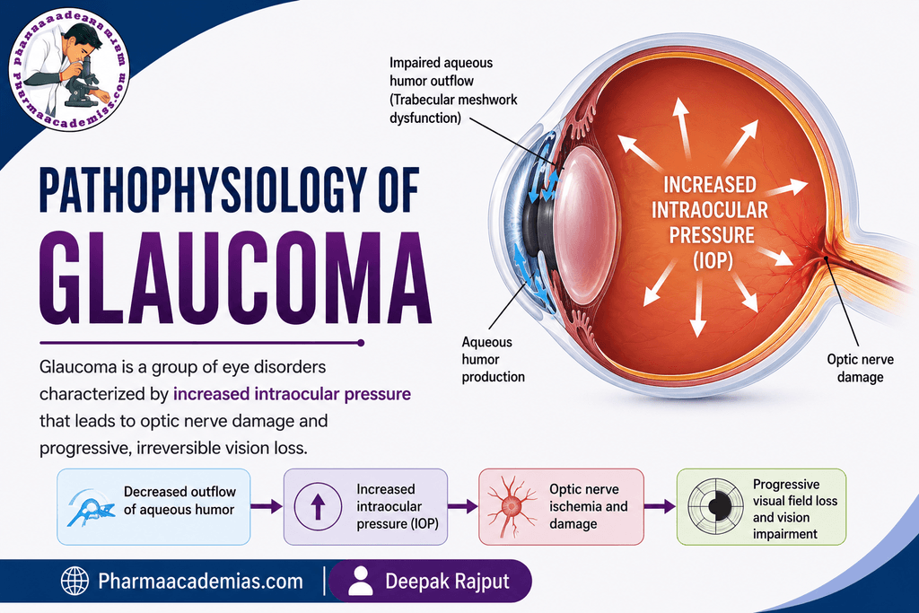
Pathophysiology of Glaucoma: Glaucoma is a group of progressive eye disorders characterized by damage to the optic nerve, particularly the retinal ganglion cell axons, leading to characteristic visual field loss. The major risk factor is often increased intraocular pressure (IOP), although glaucoma can also develop with IOP within the statistically normal range. The disease is usually chronic and progressive, and without appropriate treatment it can result in irreversible blindness.
Pathophysiology of Glaucoma
Step 1: Normal Aqueous Humor Circulation
To understand the pathophysiology of glaucoma, it is important to first understand how aqueous humor normally circulates.
Aqueous humor is a clear fluid that provides nutrients to structures such as the cornea and lens and helps maintain the shape of the eye.
It is mainly produced by the ciliary processes in the posterior chamber of the eye.
The normal pathway is:
Ciliary processes → Posterior chamber → Pupil → Anterior chamber → Trabecular meshwork → Schlemm’s canal → Episcleral veins
A smaller proportion of aqueous humor leaves the eye through the uveoscleral pathway.
Under normal conditions, the rate of aqueous humor production is approximately balanced by its drainage. This maintains IOP within a normal range.
Step 2: Disturbance in Aqueous Humor Drainage
The central event in many forms of glaucoma is reduced drainage of aqueous humor.
When aqueous humor cannot leave the eye efficiently, fluid begins to accumulate within the anterior segment.
This causes an increase in intraocular pressure.
The mechanism of impaired drainage differs according to the type of glaucoma.
Open-angle glaucoma
In primary open-angle glaucoma, the iridocorneal angle remains anatomically open, but resistance to aqueous humor outflow increases, particularly through the trabecular meshwork.
The trabecular meshwork may undergo structural and functional changes, resulting in reduced aqueous humor drainage.
Therefore:
Open angle → Increased outflow resistance → Reduced aqueous humor drainage → Increased IOP
Angle-closure glaucoma
In angle-closure glaucoma, the drainage angle becomes physically narrowed or closed.
This prevents aqueous humor from reaching the trabecular meshwork effectively.
A common mechanism involves forward movement of the iris, which obstructs the drainage angle.
Therefore:
Narrow/closed angle → Obstruction of trabecular outflow → Rapid rise in IOP
Step 3: Increase in Intraocular Pressure
When aqueous humor production exceeds its drainage, IOP increases.
Increased IOP does not automatically mean that glaucoma is present. Some individuals have elevated IOP without optic nerve damage, a condition known as ocular hypertension.
Conversely, optic nerve damage can occur even when IOP is not markedly elevated, as seen in normal-tension glaucoma.
Therefore, glaucoma is best understood as a disease involving optic nerve damage and retinal ganglion cell loss, with IOP being an important but not the only contributing factor.
Step 4: Mechanical Stress on the Optic Nerve Head
The optic nerve connects the retina to the brain.
The axons of retinal ganglion cells leave the eye through the optic nerve head, particularly through a specialized connective tissue structure called the lamina cribrosa.
When IOP becomes elevated, mechanical stress can develop at the optic nerve head.
The increased pressure may cause deformation of the lamina cribrosa and compression or distortion of retinal ganglion cell axons.
The simplified mechanism is:
↑ IOP → Mechanical stress at optic nerve head → Lamina cribrosa deformation → Axonal injury
This is one of the major mechanisms involved in glaucomatous optic neuropathy.
Step 5: Impairment of Axonal Transport
Retinal ganglion cell axons transport important substances between the cell body and the brain.
This process is called axonal transport.
Increased mechanical stress around the optic nerve head can interfere with both anterograde and retrograde axonal transport.
As a result, essential neurotrophic signals and other cellular materials may not be transported normally.
This contributes to dysfunction and eventual death of retinal ganglion cells.
Thus:
Optic nerve stress → Impaired axonal transport → Retinal ganglion cell dysfunction → Cell death
Step 6: Retinal Ganglion Cell Injury
Retinal ganglion cells (RGCs) are particularly important because their axons form the optic nerve.
Damage to these cells is a major pathological feature of glaucoma.
Initially, retinal ganglion cells may become functionally impaired. With continued injury, they undergo structural damage and eventually die.
The loss of RGCs results in progressive loss of their corresponding visual field.
Step 7: Role of Reduced Ocular Perfusion
The optic nerve head requires an adequate blood supply.
Glaucoma may involve abnormalities in ocular blood flow and vascular regulation, particularly in susceptible individuals.
Reduced ocular perfusion may cause inadequate oxygen and nutrient delivery to the optic nerve head.
This can make retinal ganglion cells more vulnerable to injury.
The process can be represented as:
Vascular dysregulation → Reduced optic nerve perfusion → Cellular stress → Retinal ganglion cell injury
This mechanism is particularly relevant in some patients with normal-tension glaucoma.
Step 8: Oxidative Stress and Mitochondrial Dysfunction
Retinal ganglion cells have high metabolic requirements.
Chronic stress associated with glaucoma can contribute to oxidative stress and mitochondrial dysfunction.
Oxidative stress occurs when the production of reactive oxygen species exceeds the body’s ability to neutralize them.
This can damage:
- Cellular membranes
- Proteins
- DNA
- Mitochondria
Mitochondrial dysfunction further reduces the ability of retinal ganglion cells to produce energy.
Therefore:
Chronic stress → Oxidative stress + mitochondrial dysfunction → Reduced cellular energy → RGC damage
Step 9: Neuroinflammation and Glial Activation
Glaucomatous optic nerve damage is also associated with changes in supporting cells, including astrocytes and microglia.
Mechanical and metabolic stress can activate these cells.
Activated glial cells can release inflammatory mediators and other molecules that may contribute to a damaging microenvironment around retinal ganglion cells.
Thus, chronic glaucoma involves not only mechanical pressure but also vascular, metabolic, inflammatory, and cellular mechanisms.
Step 10: Apoptosis of Retinal Ganglion Cells
Persistent cellular stress ultimately activates pathways leading to apoptosis, or programmed cell death.
The loss of retinal ganglion cells is progressive.
The sequence can be summarized as:
↑ IOP / vascular and cellular stress → Axonal dysfunction → RGC injury → Apoptotic signaling → RGC death
Once retinal ganglion cells and their axons are lost, they generally cannot be regenerated naturally, which explains why glaucomatous visual loss is irreversible.
Step 11: Optic Disc Changes
As retinal ganglion cell axons are progressively lost, characteristic structural changes develop at the optic nerve head.
One important finding is increased optic disc cupping.
The normal optic disc contains a central depression called the optic cup. In glaucoma, loss of neural tissue can cause the cup to become progressively enlarged relative to the disc.
This is called glaucomatous cupping.
Other structural changes may include:
- Enlargement of the cup-to-disc ratio
- Thinning of the neuroretinal rim
- Retinal nerve fiber layer thinning
- Optic disc hemorrhage in some patients
Step 12: Retinal Nerve Fiber Layer Damage
The axons of retinal ganglion cells form the retinal nerve fiber layer (RNFL).
As ganglion cells are lost, the RNFL becomes progressively thinner.
Structural RNFL damage may occur before a patient notices significant visual symptoms.
Modern glaucoma assessment therefore commonly evaluates both:
Optic nerve structure + Retinal nerve fiber layer + Visual field
Step 13: Progressive Visual Field Loss
The loss of retinal ganglion cells produces corresponding areas of visual field loss.
Early glaucoma may produce relatively subtle defects, and patients may not notice them because the disease can initially affect peripheral vision while central vision remains relatively preserved.
As the disease progresses, visual field defects become more extensive.
Advanced glaucoma can cause severe restriction of the visual field and eventually profound loss of vision.
Step 14: Advanced Glaucoma and Blindness
If glaucomatous damage continues without adequate control, progressive loss of retinal ganglion cells and optic nerve fibers occurs.
The final sequence is:
Persistent pathological process
↓
Retinal ganglion cell loss
↓
Optic nerve fiber loss
↓
Progressive optic disc cupping
↓
Visual field loss
↓
Severe visual impairment
↓
Irreversible blindness
Important Point
The simplified textbook explanation is that glaucoma occurs because increased IOP damages the optic nerve. However, the actual pathophysiology is more complex. IOP is an important modifiable risk factor, but glaucoma can occur at statistically normal IOP, and many people with elevated IOP never develop glaucomatous optic neuropathy. Susceptibility of the optic nerve, ocular perfusion, age-related changes, genetics, and cellular mechanisms also influence disease progression.
One-Line Summary
Glaucoma is a progressive optic neuropathy in which retinal ganglion cells and their axons are lost, commonly associated with impaired aqueous humor outflow and elevated IOP, resulting in optic nerve head damage, optic disc cupping, and irreversible visual field loss.
Editorial Note
This article has been carefully researched and written by Deepak Rajput with a focus on accuracy, clarity, and evidence-based healthcare information. Pharmaacademias.com