What is Neuromuscular Junction: The neuromuscular junction (NMJ) is a specialized synapse or communication site between a motor neuron and a skeletal muscle fiber. It is the point where the nervous system transmits signals to skeletal muscles, initiating muscle contraction. The neuromuscular junction plays a vital role in voluntary movements such as walking, running, writing, speaking, and lifting objects. Without a properly functioning neuromuscular junction, muscles cannot receive nerve impulses and therefore cannot contract effectively. Pharmaacademias.com
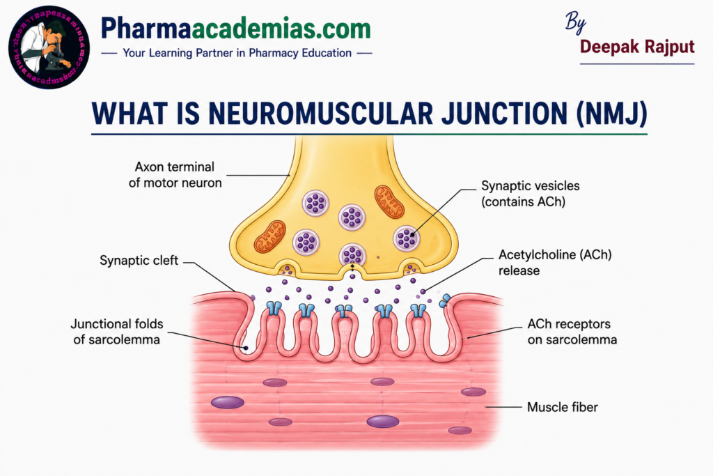
The neuromuscular junction converts an electrical signal (nerve impulse) into a chemical signal (release of a neurotransmitter) and then back into an electrical signal (muscle action potential). This process ensures rapid and efficient communication between the nervous system and skeletal muscles. The neurotransmitter responsible for this communication is acetylcholine (ACh).
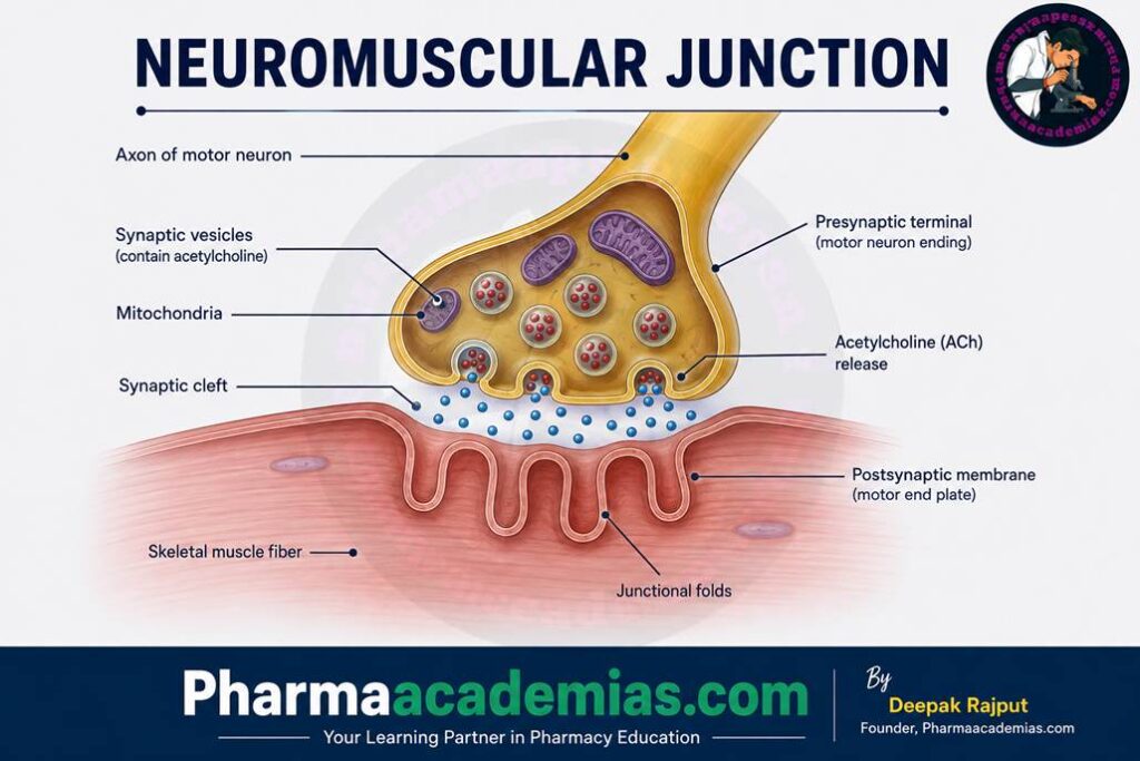
Structure of the Neuromuscular Junction
The neuromuscular junction consists of three main components:
- Presynaptic Terminal (Motor Neuron Terminal)
- Synaptic Cleft
- Postsynaptic Membrane (Motor End Plate)
Each component has a specific role in transmitting nerve impulses to the muscle.
1. Presynaptic Terminal (Motor Neuron Terminal)
The presynaptic terminal is the enlarged end of a motor neuron where the nerve impulse reaches before being transmitted to the muscle fiber. It contains numerous synaptic vesicles filled with acetylcholine (ACh), the neurotransmitter responsible for stimulating muscle contraction.
The terminal also contains voltage-gated calcium channels. When an action potential reaches the nerve ending, these channels open, allowing calcium ions (Ca²⁺) to enter the nerve terminal. The influx of calcium triggers the fusion of synaptic vesicles with the cell membrane, leading to the release of acetylcholine into the synaptic cleft by exocytosis.
In addition, the presynaptic terminal contains many mitochondria that provide ATP required for neurotransmitter synthesis, vesicle transport, and other cellular activities.
2. Synaptic Cleft
The synaptic cleft is a narrow gap, approximately 20–50 nanometers wide, that separates the motor neuron from the muscle fiber. Although extremely small, this space is essential because it allows acetylcholine to diffuse from the nerve terminal to the muscle membrane.
The synaptic cleft contains the enzyme acetylcholinesterase (AChE). This enzyme rapidly breaks down acetylcholine into acetate and choline after it has stimulated the muscle fiber. The breakdown of acetylcholine prevents continuous muscle stimulation and allows the muscle to relax when nerve impulses stop.
3. Postsynaptic Membrane (Motor End Plate)
The postsynaptic membrane, also called the motor end plate, is a specialized region of the muscle fiber’s plasma membrane (sarcolemma). It contains numerous junctional folds, which increase the surface area available for acetylcholine receptors.
The motor end plate is rich in nicotinic acetylcholine receptors (nAChRs). When acetylcholine binds to these receptors, ligand-gated ion channels open, allowing sodium ions (Na⁺) to enter the muscle fiber. This causes depolarization of the muscle membrane and generates a muscle action potential, which spreads along the sarcolemma and into the T-tubules, ultimately triggering muscle contraction.
Physiology of Neuromuscular Transmission
Transmission of nerve impulses at the neuromuscular junction occurs through a series of carefully coordinated steps.
Step 1: Arrival of the Nerve Impulse
The process begins when an action potential travels along a motor neuron and reaches the presynaptic terminal. This electrical impulse is generated in response to signals from the brain or spinal cord.
Step 2: Opening of Calcium Channels
The arrival of the action potential depolarizes the presynaptic membrane, causing voltage-gated calcium channels to open. Calcium ions rapidly enter the nerve terminal due to the concentration gradient.
The increase in intracellular calcium concentration is the key signal that initiates neurotransmitter release.
Step 3: Release of Acetylcholine
The calcium ions stimulate synaptic vesicles containing acetylcholine to fuse with the presynaptic membrane. Acetylcholine is then released into the synaptic cleft through exocytosis.
A single nerve impulse releases thousands of acetylcholine molecules, ensuring reliable stimulation of the muscle fiber.
Step 4: Binding of Acetylcholine to Receptors
Acetylcholine diffuses across the synaptic cleft within a fraction of a second and binds to nicotinic acetylcholine receptors located on the motor end plate.
The binding of acetylcholine changes the shape of the receptor, opening ion channels that allow sodium ions to enter the muscle fiber and a small amount of potassium ions to leave.
Step 5: Generation of Muscle Action Potential
The influx of sodium ions causes depolarization of the motor end plate, producing an end-plate potential. If this depolarization reaches the threshold level, it triggers a muscle action potential.
The muscle action potential spreads rapidly across the sarcolemma and into the T-tubules, leading to the release of calcium ions from the sarcoplasmic reticulum and initiating muscle contraction.
Step 6: Termination of the Signal
After acetylcholine has completed its function, acetylcholinesterase quickly hydrolyzes it into acetate and choline.
Choline is actively transported back into the presynaptic terminal, where it is reused to synthesize new acetylcholine molecules.
The rapid removal of acetylcholine ensures that the muscle contracts only when stimulated by a new nerve impulse.
Functions of the Neuromuscular Junction
The neuromuscular junction performs several essential functions:
- Transmits nerve impulses from motor neurons to skeletal muscle fibers.
- Converts electrical signals into chemical signals and then back into electrical signals.
- Initiates voluntary muscle contraction.
- Ensures rapid and efficient communication between the nervous system and muscles.
- Prevents continuous muscle stimulation through the action of acetylcholinesterase.
- Helps coordinate precise and controlled body movements.
Neurotransmitter Involved
The primary neurotransmitter at the neuromuscular junction is acetylcholine (ACh).
Its functions include:
- Binding to nicotinic acetylcholine receptors on the motor end plate.
- Opening sodium channels and producing depolarization.
- Initiating muscle action potentials.
- Triggering muscle contraction.
After its action, acetylcholine is rapidly broken down by acetylcholinesterase, allowing the muscle to relax.
Clinical Significance of the Neuromuscular Junction
The neuromuscular junction is involved in several important diseases and is also the target of certain drugs and toxins.
Myasthenia Gravis: Myasthenia gravis is an autoimmune disorder in which antibodies attack nicotinic acetylcholine receptors at the neuromuscular junction. As a result, fewer receptors are available for acetylcholine, leading to muscle weakness and fatigue. Symptoms often worsen with repeated muscle use and improve with rest.
Lambert-Eaton Myasthenic Syndrome: This disorder is caused by antibodies against presynaptic voltage-gated calcium channels. Reduced calcium entry decreases acetylcholine release, resulting in muscle weakness.
Botulism: Botulinum toxin, produced by Clostridium botulinum, blocks the release of acetylcholine from the presynaptic terminal. This prevents muscle contraction and causes flaccid paralysis. Small, controlled doses of botulinum toxin are also used therapeutically to treat muscle spasms and in cosmetic procedures.
Curare Poisoning: Curare blocks nicotinic acetylcholine receptors on the motor end plate. Although acetylcholine is released normally, it cannot bind to its receptors, preventing muscle contraction and causing paralysis.
Organophosphate Poisoning: Organophosphate compounds inhibit acetylcholinesterase, resulting in excessive accumulation of acetylcholine in the synaptic cleft. Continuous stimulation of the muscle leads to muscle twitching, spasms, weakness, and eventually paralysis if untreated.
Factors Affecting Neuromuscular Transmission
Normal neuromuscular transmission depends on several factors:
- Healthy motor neurons.
- Adequate synthesis and release of acetylcholine.
- Functional voltage-gated calcium channels.
- Intact nicotinic acetylcholine receptors.
- Normal activity of acetylcholinesterase.
- Sufficient ATP for neurotransmitter synthesis and ion transport.
- Proper electrolyte balance, particularly calcium, sodium, and potassium ions.
Summary Table
| Component | Main Function |
| Presynaptic Terminal | Releases acetylcholine in response to a nerve impulse |
| Voltage-Gated Calcium Channels | Allow calcium entry to trigger neurotransmitter release |
| Synaptic Vesicles | Store and release acetylcholine |
| Synaptic Cleft | Space through which acetylcholine diffuses |
| Acetylcholinesterase | Breaks down acetylcholine to terminate the signal |
| Motor End Plate | Contains acetylcholine receptors that initiate muscle depolarization |
| Nicotinic Acetylcholine Receptors | Bind acetylcholine and open ion channels |
| Muscle Fiber | Generates an action potential and contracts |
Conclusion
The neuromuscular junction is a highly specialized communication site that links the nervous system with skeletal muscles. It ensures that nerve impulses are efficiently transmitted to muscle fibers through the release of acetylcholine, leading to muscle contraction. The coordinated actions of the presynaptic terminal, synaptic cleft, motor end plate, calcium ions, acetylcholine, and acetylcholinesterase allow precise control of voluntary movements. Because of its critical role in movement, disorders affecting the neuromuscular junction can lead to muscle weakness, paralysis, and impaired motor function, highlighting its importance in both normal physiology and clinical medicine.
Editorial Note
This article has been carefully researched and written by Deepak Rajput with a focus on accuracy, clarity, and evidence-based healthcare information.