Skeletal system
The skeletal system, commonly known as the skeleton, forms the anatomical framework of bones and cartilage that offers structural support and shape to the human body. It acts as a protective and supportive structure for internal organs, enables bodily movement through muscle attachment, participates in blood cell production, and stores crucial minerals. The human skeleton comprises a intricate network of bones that collectively shape the body’s framework, providing both stability and mobility.
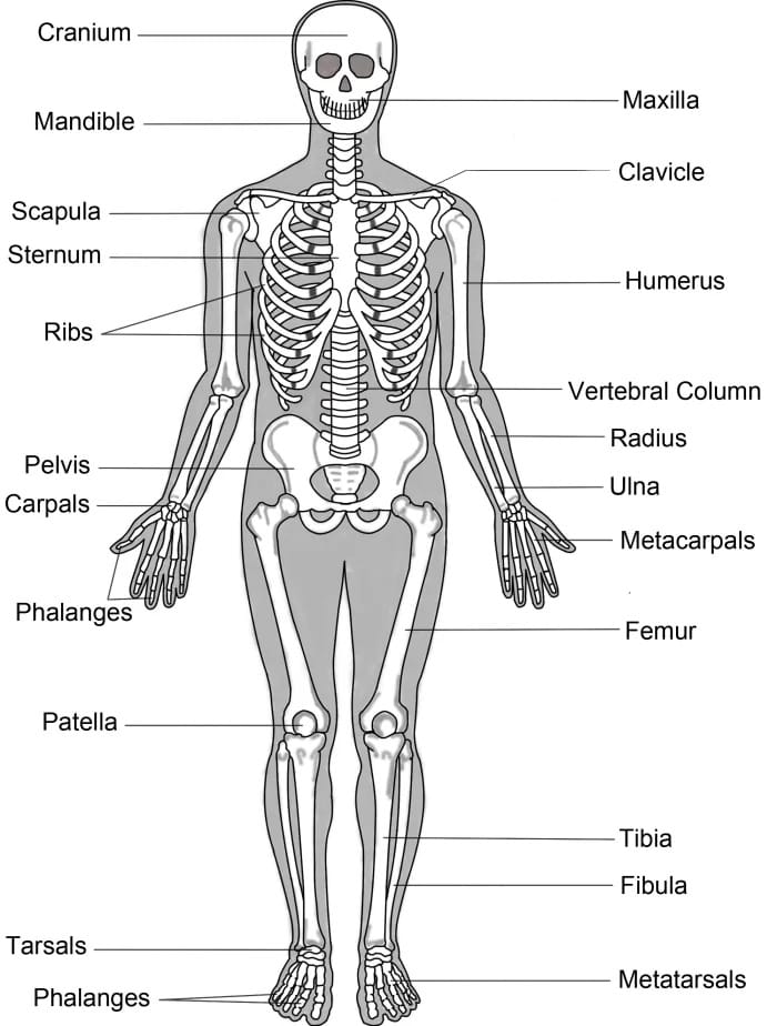
1. Axial Skeleton
The axial skeleton is the central portion of the skeletal system, primarily consisting of bones along the body’s central axis. This division includes the following components:
a. Skull: The skull forms the bony framework of the head and face in vertebrate animals, including humans. It encases and protects the brain while supporting sensory organs like the eyes, ears, and nose. The human skull, a complex structure, comprises several bones, including the cranium, which surrounds and safeguards the brain, and facial bones. It serves essential functions such as protecting the brain, anchoring facial expression-controlling muscles, and housing sensory organs responsible for vision, hearing, and olfaction.

b. Vertebral Column (Spine): The spine, also known as the vertebral column or backbone, plays a fundamental role in the human skeletal system. It provides structural support, protects the spinal cord, and enables various body movements. Composed of individual bones called vertebrae stacked on top of each other, the spine is a flexible and elongated structure. It extends from the base of the skull down to the tailbone and is divided into several regions, including the cervical (neck), thoracic (upper back), lumbar (lower back), sacral (pelvic), and coccygeal (tailbone) regions.

c. Ribs and Sternum (Breastbone): The ribcage comprises 12 ribs attached to the thoracic vertebrae. The ribs encircle and protect vital organs like the heart and lungs. The sternum is a flat bone in the front of the ribcage that provides a central anchor point for the ribs.

2. Appendicular Skeleton
The appendicular skeleton includes the bones of the limbs (appendages) and the bones that connect them to the axial skeleton. It is responsible for movement and locomotion. The appendicular skeleton is further divided into two categories:
a. Upper Limbs
Shoulder Girdle: The shoulder girdle, also known as the pectoral girdle, is a vital structural component that connects the upper limbs to the axial skeleton. It is composed of two primary bones: the clavicle (collarbone) and the scapula (shoulder blade). These bones serve as a bridge between the torso and the upper limb, ensuring stability while also allowing for a wide range of motion at the shoulder joint. The clavicle acts as a strut, holding the scapula in place and preventing it from collapsing inward, while the scapula provides attachment points for numerous muscles that facilitate the extensive movement capabilities of the arm and shoulder.
Arm: The arm consists of a single long bone, the humerus, which is positioned between the shoulder and the elbow. The humerus is the largest bone of the upper limb, and its structure plays a critical role in facilitating movement at the shoulder and elbow joints. The head of the humerus fits into the shallow cavity of the scapula, forming the ball-and-socket shoulder joint, while its distal end articulates with the bones of the forearm, the radius and ulna, forming the hinge-like elbow joint.

Forearm: The forearm is made up of two bones: the radius and the ulna. These bones are situated parallel to each other and are essential for the movement of the hand and wrist. The ulna is the larger, more medial bone, and it forms the main structural support for the elbow joint, while the radius is located on the lateral (thumb) side of the forearm and is primarily involved in wrist movement. The ability of the forearm to rotate, known as pronation and supination, is a result of the interplay between these two bones, enabling the palm of the hand to turn either upward or downward.
Hand: The human hand is a complex and finely structured part of the upper limb, allowing for intricate and highly dexterous movements. It comprises several distinct sets of bones, each contributing to its flexibility, strength, and coordination:
- Carpal Bones (eight): These are the eight bones that make up the wrist. The carpal bones are arranged in two rows, with four bones in each row. These bones are: the scaphoid, lunate, triquetrum, pisiform, trapezium, trapezoid, capitate, and hamate. They form a flexible and sturdy structure, allowing for the mobility and strength needed for various wrist movements.
- Metacarpal Bones (five): The five metacarpal bones form the structure of the palm. Each metacarpal bone corresponds to one of the fingers and serves as the foundation for the phalanges, the bones of the fingers. The metacarpals contribute to the overall shape of the hand and provide support for the fingers during various tasks.
- Phalanges (14): The phalanges are the bones that make up the fingers. There are three phalanges for each finger: the proximal, middle, and distal phalanges. However, the thumb is unique in that it has only two phalanges: the proximal and distal. These phalanges work together to enable fine motor skills, such as gripping, pinching, and manipulating objects with precision. The flexibility of the joints between these bones allows for a wide range of movement in the hand.
Together, these bones of the upper limbs provide the necessary structure and mobility to perform a multitude of tasks, from basic actions like reaching and grasping to more complex tasks requiring fine motor skills, such as writing or playing musical instruments.
b. Lower Limbs
Pelvic Girdle: The pelvic girdle, also known as the pelvic basin, is a robust bony structure that plays a crucial role in supporting the weight of the upper body when sitting and standing. It consists of two hip bones (os coxae) that are fused together to form the pelvis. Each hip bone is made up of three fused bones: the ilium, ischium, and pubis. The pelvic girdle serves as the point of attachment for the lower limbs, connecting them to the axial skeleton, and provides stability to the trunk and upper body. It also supports the organs in the lower abdomen, including the bladder, reproductive organs, and part of the intestines. The pelvis is not only crucial for weight-bearing but also contributes to mobility by providing attachment points for muscles involved in walking, running, and other activities.

Thigh: The thigh is home to the femur, which is the longest and strongest bone in the human body. The femur extends from the hip to the knee and serves as the primary bone for weight-bearing and locomotion. At its proximal end, the femur forms the hip joint with the pelvis, allowing for a wide range of motion, including walking, running, and jumping. The distal end of the femur connects with the tibia (shinbone) at the knee joint, which is a hinge joint that enables the leg to bend and straighten. The femur plays a key role in providing structural support and allowing for efficient movement during various activities, from standing to high-impact exercises.
Leg: The leg, located between the knee and the ankle, consists of two bones: the tibia and the fibula. The tibia, commonly known as the shinbone, is the larger and more robust of the two bones. It is located medially (toward the inside) in the lower leg and carries the majority of the body’s weight. The tibia is the primary bone involved in bearing the body’s load during activities such as walking, running, and standing. The fibula, on the other hand, is thinner and located laterally (toward the outside) of the tibia. While it does not bear as much weight, the fibula plays a vital role in providing lateral support and stability to the leg. It also serves as an attachment site for muscles that help stabilize the ankle and lower leg during movement.
Foot: The human foot is a highly specialized structure designed for both support and movement. It is composed of a variety of bones that work together to allow for walking, running, and balancing. The bones of the foot can be broken down into three main groups: the tarsal bones, metatarsal bones, and phalanges.
- Tarsal Bones (seven): These bones make up the rear part of the foot and the ankle, forming a sturdy and flexible structure. The seven tarsal bones include: the talus (which articulates with the tibia and fibula to form the ankle joint), the calcaneus (heel bone), the navicular, the cuboid, and the three cuneiform bones (medial, intermediate, and lateral). Together, these bones allow the foot to bear weight and absorb the shock from walking and running, while also providing the flexibility needed for proper foot movement.
- Metatarsal Bones (five): Located in the midfoot, the five metatarsal bones form the arch of the foot and connect the tarsal bones to the phalanges. These bones are numbered one through five, with the first metatarsal being the thickest and most robust as it supports the weight of the body, particularly during activities such as standing and walking. The first metatarsal, in particular, contributes to the stability of the big toe, allowing it to provide leverage and push off the ground during walking and running.
- Phalanges (14): Similar to the fingers, the toes are made up of bones called phalanges. Each toe consists of three phalanges: the proximal, middle, and distal phalanges, except for the big toe, which has only two phalanges (proximal and distal). These bones are essential for balance, grip, and movement. The toes work together to push off the ground during walking or running, and the flexibility of the phalanges allows for the foot to adapt to different surfaces and terrains. The phalanges in the toes contribute significantly to the overall function and dexterity of the foot, making it an essential component for movement and weight-bearing.
The axial and appendicular skeletons together form the complete human skeleton, providing the structural framework for the entire body. Each bone in the skeletal system has a specific function and contributes to the body’s overall mobility, support, and protection. This division of the skeletal system helps anatomists and healthcare professionals describe and study the intricate structure of the human skeleton.
Visit to: Pharmaacademias.com and Pharmacareerinsider.com