Histology of Skeletal Muscles: Skeletal muscles, also known as voluntary muscles or striated muscles, are specialized muscle tissues that are mainly attached to bones through tendons. They are responsible for producing voluntary movements of the body, such as walking, running, writing, grasping objects, maintaining posture, and making facial expressions. Skeletal muscles also contribute to the stability of joints and help maintain body posture.
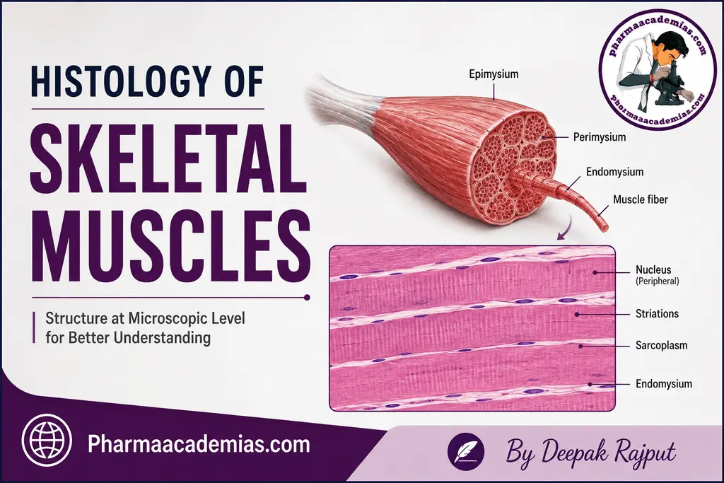
Histologically, skeletal muscle shows a highly organized arrangement of muscle fibers, myofibrils, sarcomeres, contractile proteins, and connective tissue layers. The characteristic microscopic appearance of skeletal muscle is due to the regular arrangement of actin and myosin filaments within the muscle fibers.
1. Muscle Tissue Types
There are three major types of muscle tissue in the human body:
- Skeletal muscle
- Cardiac muscle
- Smooth muscle
Skeletal Muscle Tissue
Skeletal muscle is a type of striated muscle tissue that is generally under voluntary control. The term “striated” refers to the alternating dark and light bands that can be observed under a microscope.
These striations are produced by the highly organized arrangement of actin and myosin filaments within the myofibrils. Skeletal muscle fibers are long, cylindrical, and arranged parallel to one another, allowing them to generate strong and coordinated contractions.
2. Structure of Muscle Fibers
Muscle Fiber
A muscle fiber is a single skeletal muscle cell. Unlike most ordinary cells, skeletal muscle fibers are very long and contain multiple nuclei. The nuclei are usually located at the periphery of the cell, just beneath the plasma membrane.
The plasma membrane of a skeletal muscle fiber is called the sarcolemma. It surrounds the muscle fiber and plays an important role in transmitting electrical signals that initiate muscle contraction.
Sarcoplasm
The cytoplasm of a skeletal muscle fiber is known as the sarcoplasm. It contains the normal cellular components along with specialized structures required for muscle contraction.
The sarcoplasm contains large amounts of:
- Glycogen
- Myoglobin
- Mitochondria
- Calcium ions and calcium-storage structures
Glycogen provides a readily available source of energy, while myoglobin helps store and supply oxygen to the muscle cells. The large number of mitochondria reflects the high energy requirement of skeletal muscles during contraction.
3. Myofibrils and Sarcomeres
Myofibrils
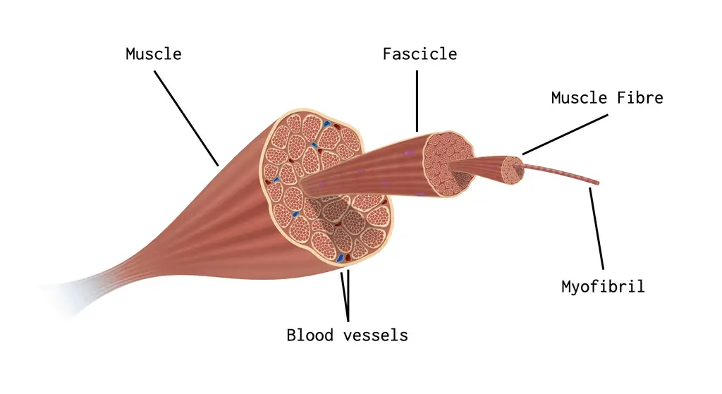
Myofibrils are long, cylindrical structures present within the sarcoplasm of skeletal muscle fibers. They extend almost the entire length of the muscle fiber.
Each muscle fiber contains numerous myofibrils arranged parallel to one another. Myofibrils are responsible for the characteristic striated appearance of skeletal muscle.
They are made up of repeating functional units called sarcomeres.
Sarcomeres
The sarcomere is the basic structural and functional unit of skeletal muscle contraction. It is the portion of a myofibril located between two successive Z-lines or Z-discs.
Each sarcomere contains two major types of protein filaments:
- Thin filaments – mainly actin
- Thick filaments – mainly myosin
The precise arrangement of these filaments produces the characteristic banding pattern observed in skeletal muscle under the microscope.
4. Actin and Myosin Filaments
Actin Filaments: Actin is the major protein of the thin filaments. These filaments are attached to the Z-line and extend toward the center of the sarcomere.
Actin works together with regulatory proteins such as tropomyosin and troponin to control muscle contraction.
Myosin Filaments: Myosin is the major protein present in the thick filaments. Myosin filaments are primarily located in the central region of the sarcomere.
Myosin molecules have specialized heads that can bind to actin and generate force during muscle contraction. The interaction between actin and myosin is essential for shortening of the sarcomere.
5. Sliding Filament Theory
The sliding filament theory explains the mechanism of skeletal muscle contraction.
When a skeletal muscle fiber receives an appropriate nerve signal, calcium ions are released inside the muscle fiber. Calcium allows the interaction between actin and myosin filaments to occur.
During contraction:
- Myosin heads attach to actin filaments.
- Myosin pulls the actin filaments toward the center of the sarcomere.
- Actin and myosin filaments slide past one another.
- The sarcomere becomes shorter.
- Shortening of many sarcomeres results in contraction of the entire muscle.
Importantly, the actin and myosin filaments themselves do not significantly shorten; instead, they slide relative to each other.
6. Connective Tissue Coverings
Skeletal muscles contain connective tissue that provides support, protection, organization, and pathways for blood vessels and nerves. Three major connective tissue layers are associated with skeletal muscle.
Endomysium: The endomysium is a thin layer of connective tissue that surrounds each individual muscle fiber. It mainly consists of delicate connective tissue and contains small blood vessels and nerve fibers that supply the muscle fiber.
Perimysium: The perimysium surrounds a group of muscle fibers. A group of muscle fibers enclosed by perimysium is called a fascicle.
The perimysium provides structural support and contains larger blood vessels and nerves that supply the muscle fibers within the fascicle.
Epimysium: The epimysium is the outermost connective tissue covering that surrounds the entire muscle. It is composed mainly of dense connective tissue.
At the ends of muscles, the connective tissue layers merge and continue into tendons, which attach skeletal muscles to bones.
7. Blood Supply and Nerve Innervation
Skeletal muscles have an extensive blood supply because muscle contraction requires a continuous supply of oxygen and nutrients.
Blood vessels present within the connective tissue layers deliver:
- Oxygen
- Glucose and other nutrients
- Hormones and other substances
They also remove carbon dioxide and metabolic waste products generated during muscle activity.
Skeletal muscles are supplied by motor nerves, which carry signals from the nervous system to the muscle fibers. These signals initiate and regulate voluntary muscle contraction.
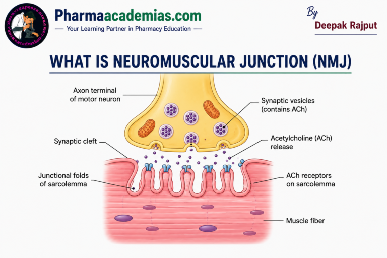
The connection between a motor neuron and a skeletal muscle fiber is called the neuromuscular junction.
8. Satellite Cells
Satellite cells are specialized stem-like cells associated with skeletal muscle fibers. They are located between the sarcolemma and the basal lamina of the muscle fiber.
Satellite cells remain relatively inactive in normal conditions but can become activated following muscle injury or during increased muscle demand.
Their major functions include:
- Muscle growth
- Muscle repair
- Regeneration of damaged muscle fibers
- Contribution to adaptation of skeletal muscle
They are therefore important for maintaining the structural integrity and regenerative capacity of skeletal muscle.
Histological Features of Skeletal Muscle
Under a light microscope, skeletal muscle can generally be identified by several characteristic features:
- Long, cylindrical and unbranched muscle fibers
- Multiple nuclei located at the periphery of the fibers
- Prominent cross-striations
- Parallel arrangement of muscle fibers
- Numerous myofibrils within each muscle fiber
- Connective tissue surrounding individual fibers and fascicles
These features help distinguish skeletal muscle from cardiac and smooth muscle tissues.
Conclusion
The histology of skeletal muscle demonstrates a highly organized arrangement of muscle fibers, myofibrils, sarcomeres, actin and myosin filaments, and connective tissue coverings. The sarcomere acts as the fundamental functional unit of contraction, while the interaction between actin and myosin produces muscle shortening according to the sliding filament theory.
The endomysium, perimysium, and epimysium provide structural support and also contain pathways for blood vessels and nerves. A rich blood supply and proper nerve innervation ensure efficient muscle function, while satellite cells contribute to muscle growth, repair, and regeneration.
Thus, the microscopic organization of skeletal muscle is closely related to its major functions, particularly voluntary movement, maintenance of posture, joint stabilization, and generation of body heat.
Editorial Note
This article has been carefully researched and written by Deepak Rajput with a focus on accuracy, clarity, and evidence-based healthcare information. Pharmaacademias.com