Formation of hemoglobin: Haemoglobin (Hb) is a complex, iron-containing protein present in the red blood cells (erythrocytes). It is responsible primarily for the transport of oxygen from the lungs to the tissues and contributes to the transport of carbon dioxide and the maintenance of acid–base balance. Haemoglobin gives blood its characteristic red colour and is one of the most important proteins for maintaining normal tissue oxygenation.
Haemoglobin is synthesized during erythropoiesis, the process of red blood cell formation that occurs mainly in the red bone marrow. Haemoglobin synthesis begins in immature erythroid precursor cells and continues as these cells develop. As erythroblasts mature, they accumulate increasing amounts of haemoglobin, eventually expel their nuclei, and develop into reticulocytes and then mature erythrocytes.
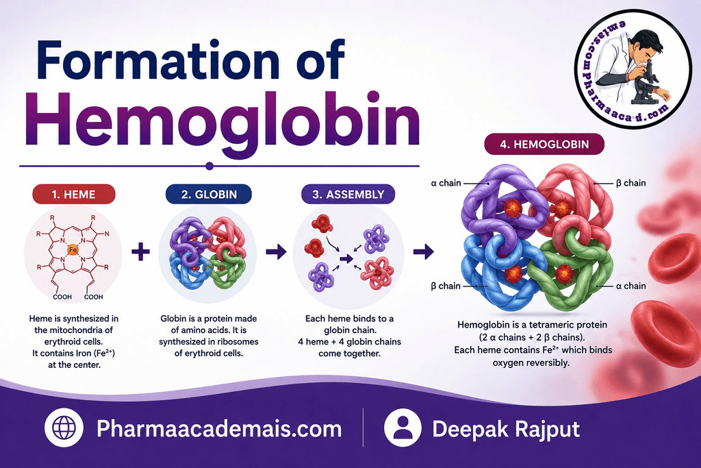
The formation of haemoglobin is a coordinated biochemical process involving the synthesis of heme and globin. Heme contains iron and is responsible for binding oxygen, whereas globin consists of protein chains that surround and support the heme groups.
A simplified representation is:
Haemoglobin = Heme + Globin
1. Structure of Haemoglobin
Normal adult haemoglobin, known as HbA, is a tetrameric protein composed of:
- Two alpha (α) globin chains
- Two beta (β) globin chains
- Four heme groups
Thus:
HbA = α₂β₂
Each globin chain is associated with one heme group, giving each haemoglobin molecule four heme groups.
Each heme group contains one atom of ferrous iron (Fe²⁺). This iron atom can bind one molecule of oxygen. Therefore, one haemoglobin molecule can reversibly bind up to four molecules of oxygen.
The globin portion provides the protein framework and influences haemoglobin’s oxygen-binding properties.
2. Main Components Required for Haemoglobin Formation
The formation of haemoglobin requires several important components:
- Iron
- Amino acids
- Glycine
- Succinyl-CoA
- Pyridoxal phosphate (vitamin B6)
- Globin genes and ribosomes
- Enzymes involved in heme synthesis
- Adequate erythropoietic activity
- Healthy bone marrow
- Adequate vitamin B12 and folate indirectly, through their roles in normal erythroid cell proliferation and maturation
Among these, iron is particularly important because it forms the central atom of the heme molecule.
3. Sites of Haemoglobin Formation
Haemoglobin is synthesized primarily in developing red blood cells in the bone marrow.
The synthesis occurs during erythropoiesis, particularly in erythroblasts and other immature erythroid cells.
During fetal development, haemoglobin synthesis occurs in the hematopoietic tissues responsible for fetal erythropoiesis, including the fetal liver and later the bone marrow.
After birth, the bone marrow becomes the major site of red blood cell and haemoglobin production.
4. Two Major Parts of Haemoglobin Synthesis
Haemoglobin synthesis can be understood as two interconnected processes:
A. Heme synthesis: This produces the iron-containing heme component.
B. Globin synthesis: This produces the protein chains that form the globin component.
Finally:
Heme + Globin → Haemoglobin
Both processes occur simultaneously within developing erythroid cells.
5. Formation of Heme
Heme is a porphyrin compound containing a central ferrous iron atom.
The synthesis of heme occurs partly in the mitochondria and partly in the cytoplasm of developing erythroid cells.
The pathway begins with:
Glycine + Succinyl-CoA
and eventually produces:
Protoporphyrin IX → Heme
Iron is then inserted into protoporphyrin IX to produce heme.
6. First Step of Heme Synthesis
The first step of heme synthesis occurs in the mitochondria.
Glycine combines with succinyl-CoA to form δ-aminolevulinic acid (ALA).
This reaction is catalyzed by:
ALA synthase (ALAS)
and requires pyridoxal phosphate, the active form of vitamin B6, as a cofactor.
The reaction can be represented as:
Glycine + Succinyl-CoA → δ-Aminolevulinic acid (ALA)
This is an important regulatory step in heme synthesis.
7. Formation of Porphobilinogen
ALA moves into the cytoplasm, where two molecules of ALA combine to form porphobilinogen (PBG).
The enzyme involved is:
ALA dehydratase
Thus:
2 ALA → Porphobilinogen
Porphobilinogen is an important intermediate in the formation of the porphyrin ring.
8. Formation of Porphyrin Precursors
Several enzymatic reactions subsequently convert porphobilinogen into progressively larger porphyrin intermediates.
The pathway proceeds through compounds including:
- Porphobilinogen
- Hydroxymethylbilane
- Uroporphyrinogen III
- Coproporphyrinogen III
- Protoporphyrinogen IX
- Protoporphyrin IX
These reactions eventually produce the porphyrin ring required for heme formation.
9. Formation of Protoporphyrin IX
The intermediate protoporphyrinogen IX is converted into protoporphyrin IX.
Protoporphyrin IX is the immediate porphyrin precursor into which iron is inserted to form heme.
This is an essential stage because the porphyrin ring must be correctly formed before iron can be incorporated.
10. Incorporation of Iron
The final major step in heme synthesis involves the insertion of ferrous iron (Fe²⁺) into protoporphyrin IX.
The enzyme responsible is:
Ferrochelatase
The reaction is:
Protoporphyrin IX + Fe²⁺ → Heme
The iron atom occupies the central position of the porphyrin ring.
This produces the functional heme group capable of reversibly binding oxygen.
11. Importance of Iron in Haemoglobin Formation
Iron is essential for haemoglobin because oxygen binds directly to the ferrous iron atom within the heme group.
The iron must be in the Fe²⁺ (ferrous) state for normal reversible oxygen binding.
Iron deficiency reduces the ability of developing erythroid cells to synthesize haemoglobin. As a result, red blood cells may become smaller and contain less haemoglobin, producing microcytic hypochromic anemia.
Therefore, adequate iron availability is essential for normal erythropoiesis and haemoglobin synthesis.
12. Globin Synthesis
The second major component of haemoglobin is globin.
Globin is a protein consisting of amino acid chains synthesized by ribosomes within developing erythroid cells.
The globin chains are encoded by specific genes.
In normal adult haemoglobin, the major globin chains are:
- α chains
- β chains
Two α chains and two β chains combine to form the globin component of HbA.
Thus:
2α + 2β → α₂β₂
13. Formation of Haemoglobin
Once heme and globin have been synthesized, they combine to form haemoglobin.
Each globin chain associates with one heme group.
Therefore:
1 globin chain + 1 heme group → haemoglobin subunit
Four such subunits assemble to form a complete haemoglobin molecule.
A normal adult HbA molecule contains:
2 α-globin chains + 2 β-globin chains + 4 heme groups
This structure enables haemoglobin to transport oxygen efficiently.
14. Haemoglobin Formation During Erythropoiesis
Haemoglobin synthesis is closely associated with the maturation of erythroid cells.
The general sequence is:
Hematopoietic stem cell
↓
Myeloid progenitor
↓
Erythroid progenitor
↓
Proerythroblast
↓
Basophilic erythroblast
↓
Polychromatophilic erythroblast
↓
Orthochromatic erythroblast
↓
Reticulocyte
↓
Mature erythrocyte
During this process, haemoglobin progressively accumulates inside the developing cell.
As haemoglobin concentration increases, the cytoplasm changes from strongly basophilic to more eosinophilic because of the increasing amount of haemoglobin.
15. Role of the Bone Marrow
The bone marrow provides the environment required for normal haemoglobin synthesis.
Developing erythroid cells are supported by the hematopoietic microenvironment and interact with other marrow cells.
Macrophages within erythroblastic islands can provide important support to developing erythroid cells.
The bone marrow also provides access to nutrients, iron, regulatory signals, and growth factors required for erythropoiesis.
16. Role of Erythropoietin
Erythropoietin (EPO) is a major hormonal regulator of red blood cell production.
It is produced predominantly by the kidneys in response to reduced oxygen availability.
The sequence can be summarized as:
Reduced tissue oxygenation
↓
Increased EPO secretion
↓
Stimulation of erythroid progenitor cells
↓
Increased erythrocyte production
↓
Increased haemoglobin-containing RBCs
↓
Improved oxygen delivery
Thus, erythropoietin indirectly promotes haemoglobin production by stimulating the production and maturation of erythroid cells.
17. Role of Iron
Iron is one of the most important nutrients required for haemoglobin synthesis.
Dietary iron is absorbed primarily in the small intestine. Once absorbed, iron is transported in the blood mainly bound to transferrin.
Developing erythroid cells obtain iron from transferrin through receptor-mediated uptake.
Inside erythroid cells, iron is incorporated into protoporphyrin IX to form heme.
Excess iron can be stored mainly as:
- Ferritin
- Hemosiderin
The body carefully regulates iron absorption and recycling because iron is essential but excessive free iron can be harmful.
18. Role of Transferrin
Transferrin is the major plasma protein responsible for transporting iron.
Iron absorbed from the intestine or released during recycling of old red blood cells is transported through the blood bound to transferrin.
Developing erythroid cells possess transferrin receptors that facilitate iron uptake.
The iron is subsequently delivered to the mitochondria, where it is incorporated into heme.
19. Role of Ferritin
Ferritin is an intracellular iron-storage protein.
It stores iron in a form that is relatively accessible and safer than free iron.
Ferritin is found in several tissues, including:
- Liver
- Bone marrow
- Spleen
- Macrophages
Serum ferritin is commonly used clinically as an indicator of body iron stores, although ferritin can also increase during inflammation.
Types of Haemoglobin
Different haemoglobin types are produced during development.
HbA: The major haemoglobin in healthy adults:
α₂β₂
HbA₂: A minor adult haemoglobin:
α₂δ₂
HbF: The major fetal haemoglobin:
α₂γ₂
These differences arise from changes in globin-chain gene expression during development.
Factors Affecting Haemoglobin Formation
Normal haemoglobin synthesis depends on several factors:
| Factor | Role in haemoglobin formation |
| Iron | Central component of heme |
| Glycine | Substrate for heme synthesis |
| Succinyl-CoA | Substrate for heme synthesis |
| Vitamin B6 | Cofactor for ALA synthase |
| Amino acids | Required for globin synthesis |
| Vitamin B12 | DNA synthesis and erythroid maturation |
| Folate | DNA synthesis and erythroid maturation |
| Erythropoietin | Stimulates erythroid production |
| Bone marrow | Primary site of erythrocyte production |
| Transferrin | Transports iron to developing erythroid cells |
| Ferritin | Stores iron |
| Healthy kidney function | Provides appropriate EPO production |
Conclusion
Haemoglobin formation is a complex and highly regulated biochemical process that occurs mainly in developing erythroid cells of the bone marrow. It requires the coordinated synthesis of two major components: heme and globin. Heme is synthesized through a series of enzymatic reactions involving glycine, succinyl-CoA, porphyrin intermediates, and iron. The final step involves incorporation of ferrous iron (Fe²⁺) into protoporphyrin IX by the enzyme ferrochelatase.
At the same time, globin chains are synthesized by ribosomes from amino acids. In normal adult haemoglobin, two α-globin chains and two β-globin chains combine with four heme groups to form HbA (α₂β₂). Each heme group contains one ferrous iron atom capable of reversibly binding oxygen, allowing a single haemoglobin molecule to carry up to four oxygen molecules.
Editorial Note
This article has been carefully researched and written by Deepak Rajput with a focus on accuracy, clarity, and evidence-based healthcare information. Pharmaacademias.com