1. Thalamus
Anatomical Location and Structure
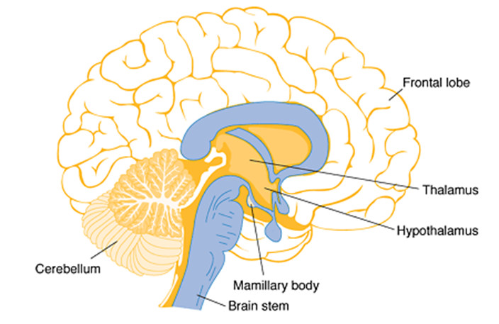
The thalamus is a large, ovoid mass of gray matter situated deep within the brain, forming the dorsal part of the diencephalon. It lies on either side of the third ventricle and is separated from each other by the narrow slit of this ventricle. Each thalamus is divided into several nuclei — such as the anterior, medial, and lateral groups, each serving specific sensory or motor functions.
Functional Overview
The thalamus acts as the main relay and integration center for sensory impulses traveling to the cerebral cortex. Almost all sensory information—except olfactory signals—passes through the thalamus before reaching the higher cortical areas for interpretation.
Key Functions
- Sensory Relay: Receives sensory input from the spinal cord and brainstem and relays it to appropriate cortical areas.
- Motor Integration: Works with the basal ganglia and cerebellum to modulate voluntary motor activities.
- Regulation of Consciousness: The thalamus plays a vital role in maintaining alertness, attention, and arousal.
- Emotion and Memory: The anterior nuclei of the thalamus are part of the limbic system, influencing emotional expression and memory processing.
Clinical Significance
Lesions or dysfunctions of the thalamus can lead to thalamic pain syndrome (Dejerine–Roussy syndrome), characterized by severe, chronic pain on the opposite side of the body. It may also cause sensory loss, motor deficits, or disturbances in consciousness.
2. Hypothalamus
Anatomical Location and Structure
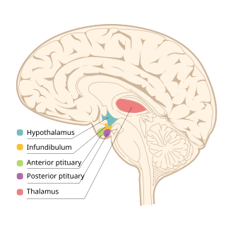
The hypothalamus lies below the thalamus and forms the floor and part of the lateral wall of the third ventricle. Despite its small size, it is one of the most vital regions of the brain. It contains several nuclei, such as the supraoptic, paraventricular, ventromedial, dorsomedial, lateral, and mammillary bodies, each with specialized functions.
Functional Overview
The hypothalamus serves as the control center for homeostasis, integrating the nervous and endocrine systems. It maintains the body’s internal environment by regulating hormonal secretion, autonomic functions, and behavioral responses.
Key Functions
- Autonomic Regulation: Controls both sympathetic and parasympathetic activities such as heart rate, blood pressure, digestion, and respiration.
- Endocrine Control: Influences the pituitary gland (hypophysis) by releasing regulatory hormones (e.g., CRH, TRH, GnRH, GHRH).
- Thermoregulation: Maintains normal body temperature via mechanisms like sweating, vasodilation, and shivering.
- Hunger and Satiety: The lateral hypothalamic area stimulates hunger, while the ventromedial nucleus signals satiety.
- Water Balance: The supraoptic and paraventricular nuclei secrete antidiuretic hormone (ADH), regulating fluid balance.
- Sleep–Wake Cycle: Works with the reticular formation and pineal gland to control circadian rhythms.
- Emotional Behavior: Influences fear, anger, pleasure, and sexual behavior through limbic connections.
Clinical Significance
Damage to the hypothalamus may result in endocrine disorders (e.g., diabetes insipidus due to lack of ADH), temperature dysregulation, obesity or anorexia, sleep disorders, or emotional instability.
3. Basal Ganglia (Basal Nuclei)
Anatomical Location and Structure
The basal ganglia are a group of subcortical gray matter nuclei located deep within each cerebral hemisphere. The major components include:
- Caudate nucleus
- Putamen
- Globus pallidus (internal and external segments)
- Subthalamic nucleus
- Substantia nigra (in the midbrain)
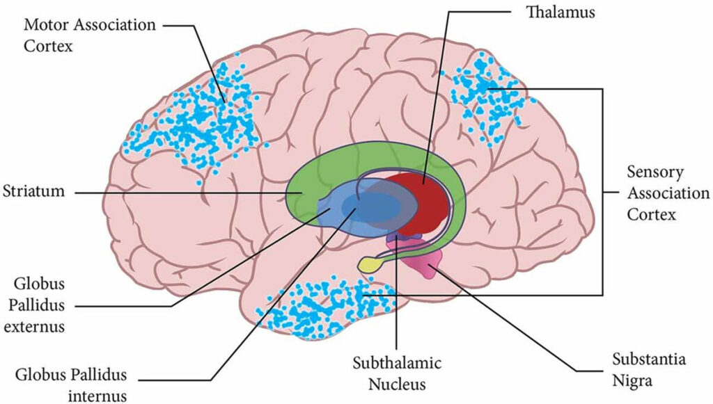
The caudate nucleus and putamen together form the striatum, which serves as the major input zone for cortical signals.
Functional Overview
The basal ganglia are primarily involved in the control of voluntary motor movements, procedural learning, habit formation, eye movements, cognition, and emotion. They form complex circuits connecting the cerebral cortex, thalamus, and brainstem, thus playing a crucial role in motor control and behavioral regulation.
Key Functions
- Motor Control: Regulate initiation, amplitude, and termination of voluntary movements.
- Posture and Muscle Tone: Help maintain smooth and coordinated muscle activity.
- Learning and Cognition: Contribute to the development of habitual and learned motor patterns.
- Reward and Motivation: The nucleus accumbens (part of the striatum) is a key player in the reward pathway, influencing motivation and addiction.
Clinical Significance
Disorders of the basal ganglia can lead to severe motor dysfunctions:
- Parkinson’s Disease: Degeneration of dopaminergic neurons in the substantia nigra, causing tremors, rigidity, and bradykinesia.
- Huntington’s Disease: Degeneration of GABAergic neurons in the caudate nucleus, leading to involuntary jerky movements (chorea).
- Hemiballismus: Damage to the subthalamic nucleus results in flinging movements of one side of the body.
Summary Table
| Structure | Location | Main Function | Clinical Significance |
|---|---|---|---|
| Thalamus | Dorsal part of diencephalon | Sensory relay, consciousness, motor integration | Thalamic pain, sensory loss |
| Hypothalamus | Below thalamus, forms floor of 3rd ventricle | Homeostasis, endocrine and autonomic control | Diabetes insipidus, obesity, thermoregulatory failure |
| Basal Ganglia | Deep within cerebral hemispheres | Motor control, habit learning, motivation | Parkinson’s, Huntington’s, dyston |