Anticoagulants are drugs or chemical substances that prevent or reduce blood coagulation, thereby inhibiting the formation of blood clots (thrombi). They are commonly used to treat or prevent conditions such as deep vein thrombosis (DVT), pulmonary embolism, and stroke in patients with atrial fibrillation or other clotting disorders. Examples include heparin, warfarin, and direct oral anticoagulants (DOACs) like rivaroxaban and apixaban.
Classification of anticoagulants
1. Anticoagulants Used In Vivo
These are anticoagulants used to prevent or treat clotting within the living body.
A. Fast Acting Anticoagulants
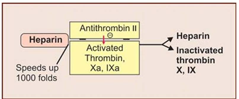
a. Heparin: Heparin is a naturally occurring anticoagulant produced by basophils and mast cells in the body. It is commonly used as a medication to prevent and treat blood clots in conditions such as deep vein thrombosis (DVT), pulmonary embolism (PE), and during surgeries or procedures that require a reduction in clotting. Heparin is available in two main forms: unfractionated heparin (UFH) and low molecular weight heparin (LMWH).
Mechanism of Action
The anticoagulant effect of heparin is primarily achieved through its interaction with antithrombin III (AT III), a natural inhibitor of several enzymes in the coagulation system. The mechanism can be detailed as follows:
Binding to Antithrombin III (AT III): Heparin binds to AT III, causing a conformational change in AT III that significantly enhances its inhibitory activity.
Inhibition of Thrombin and Factor Xa: This enhanced AT III can more effectively inhibit thrombin (Factor IIa) and Factor Xa, two critical proteases in the coagulation cascade. Thrombin is essential for converting fibrinogen to fibrin, the structural basis of a blood clot. Unfractionated heparin (UFH) can inhibit both thrombin and Factor Xa equally because of its longer polysaccharide chains. Low molecular weight heparin (LMWH) predominantly inhibits Factor Xa due to its shorter chains but has a reduced ability to inhibit thrombin.
Additional Effects:
Heparin also inactivates other factors such as IXa, XIa, and XIIa, contributing to its anticoagulant properties.
It prevents the formation and extension of clots but does not break down existing clots (a process known as fibrinolysis).
b. Low Molecular Weight Heparins (LMWHs)
Examples: Enoxaparin, Dalteparin
Enoxaparin: Enoxaparin is a low molecular weight heparin (LMWH) derived from unfractionated heparin. It is used to prevent and treat blood clots, similar to heparin but with distinct pharmacokinetic and pharmacodynamic properties. Enoxaparin is often preferred for its predictable anticoagulant response and ease of use.
Mechanism of Action
Enoxaparin works by enhancing the activity of antithrombin III (AT III), which in turn inhibits key factors in the blood coagulation process, primarily Factor Xa and to a lesser extent, thrombin (Factor IIa).
Binding to Antithrombin III (AT III): Enoxaparin binds to AT III, inducing a conformational change that significantly increases the inhibitory effect of AT III on coagulation factors.
Inhibition of Factor Xa: Enoxaparin predominantly inhibits Factor Xa. By inhibiting Factor Xa, enoxaparin prevents the conversion of prothrombin to thrombin, which is crucial for clot formation.
Partial Inhibition of Thrombin: Due to its shorter polysaccharide chains compared to unfractionated heparin, enoxaparin has a reduced ability to inhibit thrombin directly. Its primary action remains the inhibition of Factor Xa.
Dalteparin: Dalteparin is a low molecular weight heparin (LMWH) used to prevent and treat blood clots. Like other LMWHs, it is derived from unfractionated heparin and is characterized by its predictable pharmacokinetic profile and ease of use. Dalteparin is commonly used in various clinical settings to manage thromboembolic disorders.
Mechanism of Action
Dalteparin exerts its anticoagulant effects through its interaction with antithrombin III (AT III), enhancing the inhibition of specific coagulation factors.
Binding to Antithrombin III (AT III): Dalteparin binds to AT III, causing a conformational change that increases the inhibitory activity of AT III.
Inhibition of Factor Xa: The primary action of dalteparin is the inhibition of Factor Xa. By inhibiting Factor Xa, dalteparin prevents the conversion of prothrombin to thrombin, a crucial step in the formation of a blood clot.
Partial Inhibition of Thrombin: Due to its shorter polysaccharide chains compared to unfractionated heparin, dalteparin has a reduced ability to inhibit thrombin directly.
c. Heparinoids
Heparinoids are a class of medications that are chemically related to heparin. They are used for their anticoagulant and anti-inflammatory properties. Heparinoids are glycosaminoglycans, similar in structure to heparin, which is a naturally occurring anticoagulant found in the body.
Heparan Sulphate: Heparan sulfate is a glycosaminoglycan found on cell surfaces and in the extracellular matrix. It consists of repeating disaccharide units of glucosamine and uronic acid, variably sulfated, and plays key roles in cell signaling, coagulation, and development.
Mechanism of Action
Heparan sulfate functions through its interactions with various proteins, influenced by its sulfation patterns:
Binding to Antithrombin III (AT III): Heparan sulfate binds to AT III, causing a conformational change that enhances AT III’s inhibition of thrombin (Factor IIa) and Factor Xa, helping to regulate blood coagulation.
Interaction with Growth Factors and Receptors: Heparan sulfate binds to growth factors, cytokines, and cell surface receptors, modulating cell signaling pathways, which are crucial for cell growth, differentiation, and migration.
Dextran Sulphate: Dextran sulfate is a sulfated polysaccharide derived from dextran, which is a complex branched glucan (sugar polymer). It is composed of glucose molecules linked predominantly by α-1,6 glycosidic bonds, with varying degrees of sulfation. Dextran sulfate is used in various biomedical applications, including as an anticoagulant and antiviral agent.
Mechanism of Action
Dextran sulfate exerts its effects through multiple mechanisms, primarily related to its highly negatively charged sulfate groups:
Anticoagulant Action:
Binding to Antithrombin III (AT III): Dextran sulfate binds to AT III, enhancing its ability to inhibit thrombin (Factor IIa) and Factor Xa, similar to heparin. This inhibits the coagulation cascade, preventing blood clot formation.
Interference with Platelet Aggregation: Dextran sulfate can inhibit platelet aggregation by interfering with the platelet surface receptors, reducing the risk of clot formation.
Antiviral Action:
Inhibition of Viral Attachment and Entry: Dextran sulfate can bind to viral particles and host cell surfaces, preventing viruses from attaching to and entering host cells. This is particularly effective against viruses that utilize heparan sulfate proteoglycans as receptors.
Inhibition of Viral Replication: By binding to viral proteins, dextran sulfate can inhibit viral replication processes within host cells.
Danaparoid: Danaparoid is a glycosaminoglycan mixture that consists of three components: heparan sulfate, dermatan sulfate, and chondroitin sulfate. It is used as an anticoagulant and is especially noted for its use in patients who have developed heparin-induced thrombocytopenia (HIT), a condition where heparin causes an immune reaction leading to a low platelet count and increased clotting risk.
Mechanism of Action
Danaparoid exerts its anticoagulant effects through several mechanisms:
Interaction with Antithrombin III (AT III): Danaparoid enhances the activity of AT III, a natural inhibitor of coagulation factors. This leads to increased inhibition of Factor Xa and, to a lesser extent, thrombin (Factor IIa), reducing clot formation.
Inhibition of Factor Xa: Similar to heparin, danaparoid primarily inhibits Factor Xa. By inhibiting Factor Xa, danaparoid prevents the conversion of prothrombin to thrombin, thus reducing the formation of fibrin clots.
Binding and Inhibition of Other Coagulation Factors: Danaparoid also has some effect on other factors involved in the coagulation cascade due to its complex mixture of glycosaminoglycans, but its primary anticoagulant effect is through AT III-mediated inhibition of Factor Xa.
Lepirudin: Lepirudin is a direct thrombin inhibitor used as an anticoagulant, primarily in patients who have developed heparin-induced thrombocytopenia (HIT) or in situations where other anticoagulants are not suitable. It is a recombinant form of hirudin, a naturally occurring anticoagulant derived from leeches.
Mechanism of Action
Lepirudin exerts its anticoagulant effects through direct inhibition of thrombin (Factor IIa):
Direct Inhibition of Thrombin: Lepirudin binds directly to thrombin, inhibiting its activity. Thrombin is a key enzyme in the coagulation cascade responsible for converting fibrinogen to fibrin, thus preventing clot formation.By binding to both the active site of thrombin and the exosite on the thrombin molecule, lepirudin effectively prevents thrombin from catalyzing the conversion of fibrinogen and from activating factors V, VIII, and XI.
Inhibition of Fibrin Formation: Since thrombin is crucial for fibrin formation, lepirudin’s inhibition of thrombin prevents the formation of fibrin clots, reducing the risk of thrombus formation.
B. Slow Acting Anticoagulants (Oral Anticoagulants)
a. Coumarin Derivatives
Bishydroxycoumarin: Bishydroxycoumarin is a coumarin derivative and an anticoagulant. It is primarily used in the treatment and prevention of thromboembolic disorders by inhibiting the synthesis of vitamin K-dependent clotting factors.
Mechanism of Action
Bishydroxycoumarin works by inhibiting the synthesis of vitamin K-dependent clotting factors, which are essential for the coagulation cascade:
Inhibition of Vitamin K Epoxide Reductase: Bishydroxycoumarin inhibits the enzyme vitamin K epoxide reductase, which is crucial for recycling vitamin K in its active form. Vitamin K is essential for the synthesis of clotting factors II, VII, IX, and X in the liver.By inhibiting this enzyme, bishydroxycoumarin reduces the availability of active vitamin K, thereby decreasing the synthesis of these clotting factors.
Reduction in Clotting Factor Synthesis: With less vitamin K available, the synthesis of functional clotting factors is impaired, leading to a reduction in the blood’s ability to form clots.
Warfarin Sodium: Warfarin sodium is an oral anticoagulant used to prevent and treat thromboembolic disorders. It is one of the most widely used anticoagulants and works by inhibiting the synthesis of vitamin K-dependent clotting factors.
Mechanism of Action
Warfarin sodium exerts its anticoagulant effect through the inhibition of vitamin K epoxide reductase, an enzyme crucial for the regeneration of active vitamin K:
Inhibition of Vitamin K Epoxide Reductase: Warfarin sodium inhibits vitamin K epoxide reductase, which is necessary for converting vitamin K epoxide back to its active form. Active vitamin K is required for the synthesis of clotting factors II, VII, IX, and X in the liver.
Reduction of Vitamin K-Dependent Clotting Factors: By inhibiting this enzyme, warfarin sodium reduces the levels of functional vitamin K. This leads to a decreased synthesis of vitamin K-dependent clotting factors, impairing the coagulation cascade and reducing the blood’s ability to form clots.
Nicoumalone: Nicoumalone is an oral anticoagulant that belongs to the coumarin class of drugs. It is used to prevent and treat thromboembolic disorders by inhibiting vitamin K-dependent clotting factors.
Mechanism of Action
Nicoumalone exerts its anticoagulant effect similarly to warfarin and other coumarin derivatives by inhibiting the synthesis of vitamin K-dependent clotting factors:
Inhibition of Vitamin K Epoxide Reductase: Nicoumalone inhibits the enzyme vitamin K epoxide reductase. This enzyme is crucial for regenerating active vitamin K from its epoxide form.
Reduction of Vitamin K-Dependent Clotting Factors: By inhibiting vitamin K epoxide reductase, nicoumalone reduces the availability of active vitamin K. As a result, the synthesis of functional vitamin K-dependent clotting factors (Factors II, VII, IX, and X) in the liver is decreased.
Impairment of Coagulation Cascade: With lower levels of these clotting factors, the coagulation cascade is impaired, which reduces the blood’s ability to form clots.
Mechanism: Inhibit vitamin K epoxide reductase, reducing the synthesis of vitamin K-dependent clotting factors II, VII, IX, and X.
b. Indandione Derivatives
Phenindione: Phenindione is an oral anticoagulant that belongs to the indanedione class of drugs. It is used to prevent and treat thromboembolic disorders by inhibiting the synthesis of vitamin K-dependent clotting factors.
Mechanism of Action
Phenindione acts similarly to coumarin derivatives like warfarin by interfering with vitamin K metabolism:
Inhibition of Vitamin K Epoxide Reductase: Phenindione inhibits vitamin K epoxide reductase, the enzyme responsible for converting vitamin K epoxide to its active form. This process is essential for synthesizing vitamin K-dependent clotting factors.
Reduction of Vitamin K-Dependent Clotting Factors: By inhibiting this enzyme, phenindione decreases the availability of active vitamin K. This leads to reduced synthesis of vitamin K-dependent clotting factors (Factors II, VII, IX, and X) in the liver.
Impairment of Coagulation Cascade: Lower levels of these clotting factors impair the coagulation cascade, reducing the blood’s ability to form clots.
Diphenadione: Diphenadione is an oral anticoagulant that is part of the indanedione class of drugs, similar to phenindione. It is used to prevent and treat thromboembolic disorders by inhibiting the synthesis of vitamin K-dependent clotting factors.
Mechanism of Action
Diphenadione acts through a mechanism similar to other vitamin K antagonists:
Inhibition of Vitamin K Epoxide Reductase: Diphenadione inhibits the enzyme vitamin K epoxide reductase, which is essential for regenerating active vitamin K from its epoxide form.
Reduction of Vitamin K-Dependent Clotting Factors: By inhibiting this enzyme, diphenadione reduces the availability of active vitamin K. This results in decreased synthesis of vitamin K-dependent clotting factors (Factors II, VII, IX, and X) in the liver.
Impairment of Coagulation Cascade: With lower levels of these clotting factors, the coagulation cascade is impaired, leading to a reduced ability of blood to form clots.
2. Anticoagulants Used In Vitro
These are anticoagulants used to prevent blood clotting in laboratory settings, such as during blood collection or storage.
Heparin: Heparin is a naturally occurring anticoagulant that prevents blood clot formation by enhancing the activity of antithrombin III, which inhibits thrombin and factor Xa in the coagulation cascade. It is widely used in the prevention and treatment of thromboembolic disorders such as deep vein thrombosis (DVT), pulmonary embolism, and during surgeries like cardiac bypass.
Mechanism of Action of Heparin
Heparin exerts its anticoagulant effect by enhancing the activity of antithrombin III (AT-III), a natural inhibitor of coagulation factors. This leads to the inhibition of thrombin (Factor IIa) and Factor Xa, which are crucial for blood clot formation.
Stepwise Mechanism:
Binding to Antithrombin III (AT-III): Heparin binds to AT-III, inducing a conformational change that significantly increases its ability to inhibit clotting factors.
Inhibition of Thrombin (Factor IIa) and Factor Xa: Unfractionated Heparin (UFH): Inhibits both thrombin (Factor IIa) and Factor Xa.
Low Molecular Weight Heparin (LMWH): Primarily inhibits Factor Xa, with less effect on thrombin.
Prevention of Fibrin Clot Formation: By inhibiting thrombin, heparin prevents the conversion of fibrinogen into fibrin, which is essential for clot formation.
No Direct Fibrinolytic Activity: Heparin does not break down existing clots but prevents new clot formation and allows the body’s natural fibrinolytic system to dissolve clots over time.
Citrates and Oxalates: Citrates are salts or esters of citric acid that act as anticoagulants by chelating calcium ions (Ca²⁺), preventing blood clotting. Oxalates are salts of oxalic acid that inhibit blood coagulation by forming an insoluble complex with calcium, making it unavailable for the clotting process.
Mechanism of Action of Citrates and Oxalates as Anticoagulants
1. Citrates: Citrates act as calcium chelators, binding to free calcium ions (Ca²⁺) in the blood. Since calcium is essential for the activation of clotting factors in the coagulation cascade, its removal prevents blood clot formation. This process is reversible, meaning that adding calcium back can restore normal coagulation.
2. Oxalates: Oxalates inhibit coagulation by forming insoluble calcium oxalate complexes, which irreversibly remove calcium from the blood. Without free calcium, clotting factors cannot be activated, and coagulation is inhibited. This action is irreversible, unlike citrate, which allows calcium restoration.
Sodium Edetate (EDTA): Sodium edetate (EDTA) is a chelating agent that binds to metal ions, particularly calcium (Ca²⁺) and magnesium (Mg²⁺), preventing their participation in biochemical and coagulation processes.
Mechanism of Action: Sodium EDTA acts as an anticoagulant by chelating calcium ions (Ca²⁺), which are essential for the coagulation cascade.By removing free calcium, it prevents blood clotting and is commonly used in laboratory blood collection (lavender-top tubes) for hematological tests.