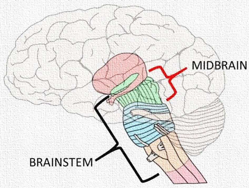
The midbrain, also known as the mesencephalon, is the uppermost part of the brainstem, positioned between the pons below and the diencephalon (thalamus and hypothalamus) above. Despite being one of the smaller regions of the brainstem, it is a vital conduit and integration center for sensory and motor pathways. The midbrain plays an essential role in vision, hearing, motor control, alertness, temperature regulation, and reflexive movements of the eyes and head.
I. Anatomy of the Midbrain
A. Location and General Structure
The midbrain forms the shortest and most superior segment of the brainstem, approximately 2.5 cm in length. It connects the forebrain (cerebrum and diencephalon) with the hindbrain (pons and cerebellum), acting as a crucial communication bridge between higher and lower neural centers.
It surrounds a narrow canal called the cerebral aqueduct (aqueduct of Sylvius), which connects the third ventricle (in the diencephalon) with the fourth ventricle (in the pons and medulla).
The midbrain is divided into two major regions by the cerebral aqueduct:
- Tectum (Dorsal Part) – located posterior to the aqueduct.
- Cerebral Peduncles (Ventral Part) – located anterior to the aqueduct and form the main bulk of the midbrain.
B. External Features
- On the Dorsal Surface (Tectum):
- The tectum contains four rounded elevations called the corpora quadrigemina:
- Superior Colliculi (2): Associated primarily with visual reflexes.
- Inferior Colliculi (2): Involved in auditory reflexes and sound localization.
- These structures collectively form the quadrigeminal plate.
- The tectum contains four rounded elevations called the corpora quadrigemina:
- On the Ventral Surface:
- Two large, rope-like structures known as the cerebral peduncles are visible. Each peduncle is divided into:
- Crus cerebri (basis pedunculi): Contains descending motor fibers from the cerebral cortex.
- Tegmentum: Contains ascending sensory tracts and nuclei of cranial nerves.
- The interpeduncular fossa lies between the peduncles and contains the oculomotor nerve (cranial nerve III) emerging from its anterior aspect.
- Two large, rope-like structures known as the cerebral peduncles are visible. Each peduncle is divided into:
C. Internal Structure
The midbrain, in cross-section, consists of three distinct regions:
1. Tectum (Roof)
- Located posterior to the cerebral aqueduct.
- Contains:
- Superior colliculi: Control visual reflexes such as tracking moving objects and coordinating head and eye movements.
- Inferior colliculi: Function as relay stations for auditory information traveling from the inner ear to the auditory cortex.
2. Tegmentum (Core of the Midbrain)
- The central portion, situated between the tectum and the crus cerebri.
- Contains several important nuclei and tracts:
- Red Nucleus: A large, reddish structure involved in motor coordination. It receives input from the cerebellum and cerebral cortex and projects to the spinal cord via the rubrospinal tract.
- Substantia Nigra: A darkly pigmented band of gray matter containing dopamine-producing neurons. It plays a key role in modulating motor activity through connections with the basal ganglia. Degeneration of this nucleus is characteristic of Parkinson’s disease.
- Cranial Nerve Nuclei:
- Oculomotor nucleus (Cranial Nerve III) – controls most of the eye’s movements.
- Trochlear nucleus (Cranial Nerve IV) – controls the superior oblique muscle of the eye.
- Reticular Formation: Part of the brainstem’s reticular activating system (RAS), which regulates arousal, consciousness, and attention.
- Ascending and Descending Tracts: The tegmentum serves as a pathway for both sensory and motor fibers connecting higher and lower centers.
3. Crus Cerebri (Basis Pedunculi)
- Located on the ventral side; composed mainly of descending motor fibers:
- Corticospinal Tract: Conducts impulses from the motor cortex to spinal motor neurons, controlling voluntary movements.
- Corticobulbar Tract: Carries impulses to motor nuclei of cranial nerves.
- Corticopontine Fibers: Connect the cerebral cortex to the pons and cerebellum for motor coordination.
II. Physiology of the Midbrain
The midbrain serves as a relay, coordination, and reflex center integrating sensory and motor pathways and controlling several vital reflexes.
A. Sensory Functions
- Visual Reflexes (Superior Colliculi):
- The superior colliculi receive input from the retina and visual cortex and coordinate reflexive eye movements.
- They are responsible for:
- Tracking moving objects.
- Pupil constriction and dilation.
- Turning the head and eyes toward visual stimuli (e.g., bright light).
- Auditory Reflexes (Inferior Colliculi):
- The inferior colliculi act as relay centers in the auditory pathway, receiving impulses from the cochlear nuclei.
- They mediate reflex responses to sound, such as turning the head toward a sudden noise (the startle reflex).
B. Motor Functions
- Substantia Nigra and Red Nucleus:
- The substantia nigra releases dopamine, which regulates voluntary motor control through its influence on the basal ganglia.
- The red nucleus assists in coordination of limb movements, especially flexor muscle tone, through the rubrospinal tract.
- Together, these nuclei ensure smooth execution of movement, posture maintenance, and muscle tone control.
- Cerebral Peduncles:
- The corticospinal, corticopontine, and corticobulbar tracts within the peduncles transmit motor commands from the cerebral cortex to lower centers, thus connecting higher cognitive control with actual motor performance.
C. Reflex and Autonomic Functions
- The midbrain is involved in several reflex activities, including:
- Pupillary reflex – constriction of the pupil in response to light.
- Accommodation reflex – adjustment of the lens for near vision.
- Startle reflex – involuntary reaction to sudden auditory or visual stimuli.
- The reticular formation of the midbrain contributes to autonomic regulation, influencing heart rate, respiration, and general arousal.
D. Regulation of Arousal and Consciousness
- The reticular activating system (RAS) within the midbrain is responsible for maintaining alertness and wakefulness.
- It filters sensory input to prevent sensory overload and ensures that important stimuli reach the conscious level.
- Damage to this system may lead to coma or loss of consciousness.
III. Blood Supply
The midbrain is supplied by branches of:
- Posterior cerebral artery
- Superior cerebellar artery
- Basilar artery
Compromise of these vessels may result in midbrain syndromes such as:
- Weber’s syndrome: Lesion in the cerebral peduncle causing contralateral paralysis and ipsilateral oculomotor palsy.
- Benedikt’s syndrome: Involvement of red nucleus leading to tremor, ataxia, and oculomotor paralysis.
IV. Clinical Correlations
| Condition | Description / Effect |
|---|---|
| Parkinson’s Disease | Degeneration of dopaminergic neurons in the substantia nigra causing tremor, rigidity, and bradykinesia. |
| Weber’s Syndrome | Damage to corticospinal fibers and oculomotor nerve causing contralateral hemiplegia and ipsilateral eye paralysis. |
| Benedikt’s Syndrome | Lesion involving red nucleus causing tremors, involuntary movements, and cranial nerve III palsy. |
| Coma or Unresponsiveness | Lesion in the reticular formation impairs arousal mechanisms. |
V. Summary Table
| Feature | Description |
|---|---|
| Location | Between pons and diencephalon |
| Major Parts | Tectum, Tegmentum, Crus Cerebri |
| Key Structures | Superior & Inferior Colliculi, Substantia Nigra, Red Nucleus, Cerebral Peduncles |
| Cranial Nerves | Oculomotor (III) and Trochlear (IV) |
| Functions | Visual and auditory reflexes, motor coordination, arousal, pupil regulation |
| Main Neurotransmitter | Dopamine (from Substantia Nigra) |
VI. Conclusion
The midbrain serves as a pivotal control center within the brainstem, integrating sensory input with motor output to generate reflexive and voluntary responses. It acts as a vital conduit between the higher brain centers and the spinal cord, while also maintaining essential functions such as alertness, vision, hearing, and motor regulation. Its structures—especially the substantia nigra, red nucleus, and colliculi—are indispensable for coordinated, adaptive, and purposeful behavior.
In summary, the midbrain exemplifies the seamless collaboration between structure and function, ensuring the harmony of movement, perception, and consciousness that defines human neural activity.