What are Tissues: The human body is an extraordinarily organized and complex structure composed of millions of cells. These cells do not function independently; instead, they group together in an organized manner to perform specialized activities essential for life. A tissue can therefore be defined as a group of structurally similar cells, along with their intercellular substances, that work collectively to perform a particular function. The branch of science that deals with the microscopic study of tissues is known as Histology.
Tissues are fundamental organizational units of the body because they form organs, and organs further combine to form organ systems. Each tissue possesses a unique structure that enables it to perform specific physiological roles. The relationship between structure and function is extremely important in understanding tissues. For example, tissues involved in protection are structurally different from tissues specialized for movement or impulse conduction.
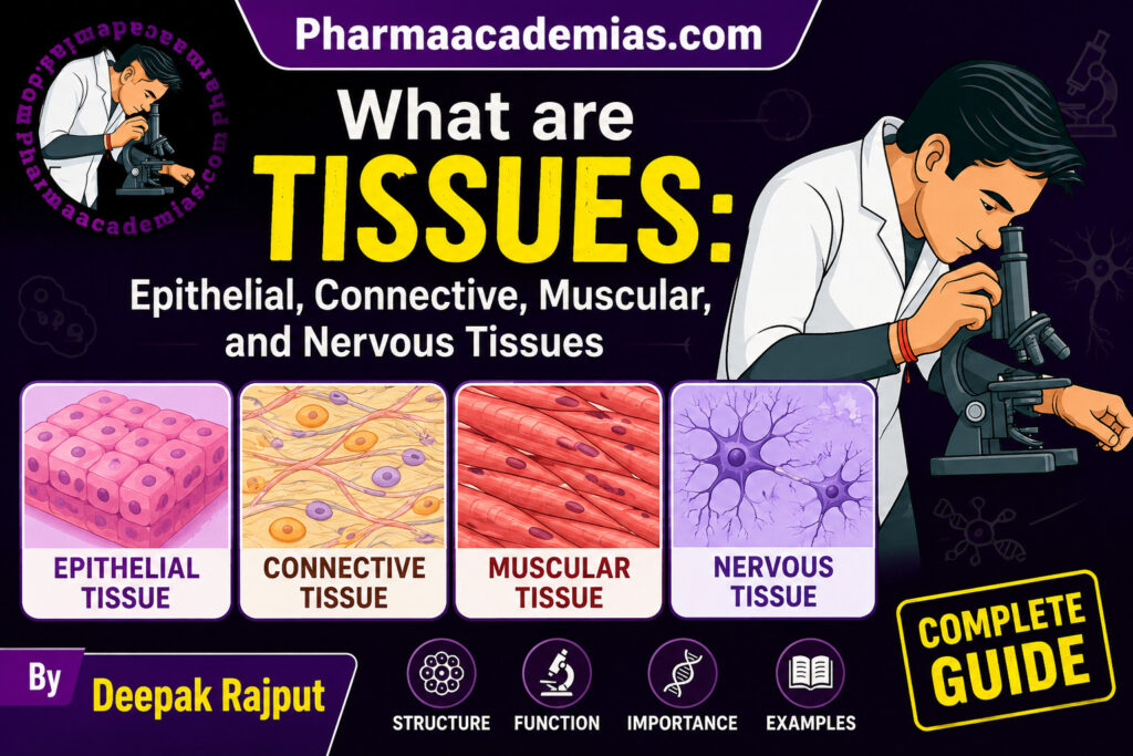
In the human body, tissues are broadly classified into four major categories: epithelial tissue, connective tissue, muscular tissue, and nervous tissue. These four tissue types collectively form the framework of all body organs and systems. Although they are distinct in structure and function, they work in close coordination to maintain homeostasis and ensure the survival of the organism.
Classification of Tissues
A. Epithelial Tissue
Epithelial tissue is one of the most important and widely distributed tissues in the body. It forms the protective covering over the external surface of the body and also lines the internal cavities, hollow organs, ducts, and blood vessels. In addition to covering and lining surfaces, epithelial tissue also gives rise to glands responsible for secretion.
Epithelial tissue acts as the body’s first line of defense against physical injury, microbial invasion, dehydration, and harmful chemicals. Besides protection, it is involved in several vital physiological functions such as absorption, secretion, filtration, diffusion, excretion, and sensory perception.
The cells of epithelial tissue are tightly packed with very little intercellular material between them. This compact arrangement forms continuous sheets that effectively cover body surfaces. The tissue rests on a thin, non-cellular structure known as the basement membrane, which anchors it to the underlying connective tissue.
An important feature of epithelial tissue is that it lacks blood vessels. Nutrients and oxygen reach epithelial cells through diffusion from nearby connective tissues. Because epithelial tissues are frequently subjected to wear and tear, they possess a remarkable ability to regenerate rapidly.
Classification of Epithelial Tissue
Epithelial tissue is mainly classified on the basis of the number of cell layers and the shape of the cells. Broadly, it is divided into simple epithelium, stratified epithelium, and glandular epithelium.
Simple Epithelium
Simple epithelium consists of a single layer of cells resting directly on the basement membrane. Since the tissue is thin, it is primarily involved in activities such as absorption, secretion, filtration, and diffusion.
Simple Squamous Epithelium
Simple squamous epithelium is composed of a single layer of extremely thin, flat, polygonal cells. The cells resemble floor tiles when viewed from above. Each cell contains a flattened central nucleus. Because the cells are very thin, this epithelium forms delicate membranes that allow substances to pass easily across them.
This type of epithelium is ideally suited for diffusion and filtration. It is found in areas where rapid exchange of substances occurs. For example, the alveoli of the lungs are lined by simple squamous epithelium, allowing efficient exchange of oxygen and carbon dioxide between air and blood. Similarly, it forms the lining of blood vessels, where it is known as endothelium, providing a smooth surface for blood circulation. It also lines body cavities as mesothelium and participates in secretion of lubricating fluid.
The primary function of simple squamous epithelium is to facilitate diffusion, osmosis, filtration, and smooth movement of fluids.
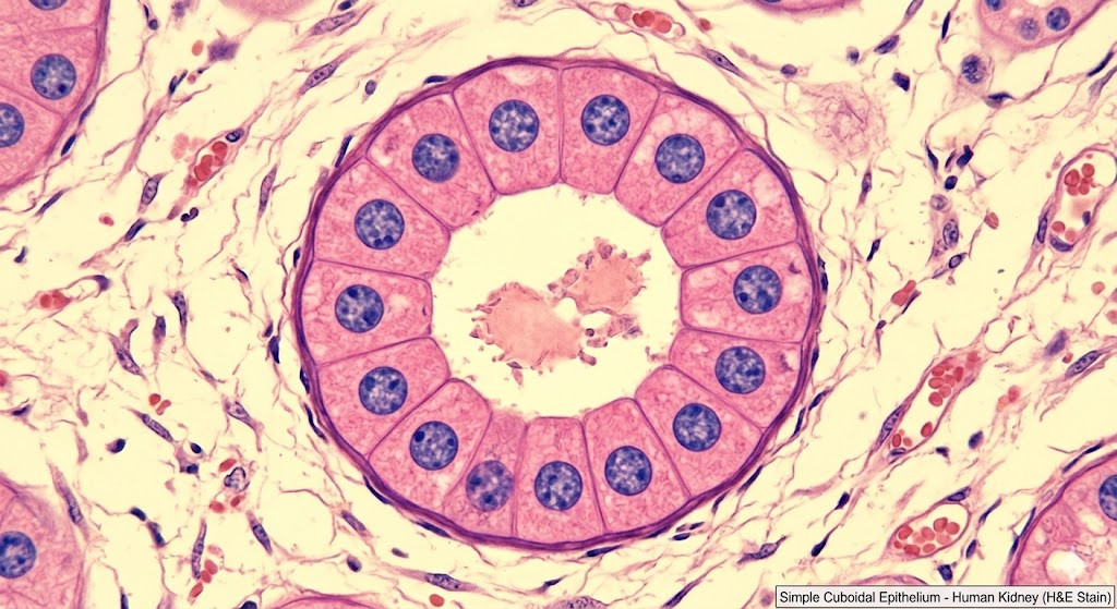
Simple Cuboidal Epithelium
Simple cuboidal epithelium consists of cube-shaped cells arranged in a single layer. The nuclei are generally spherical and located centrally within the cells. Compared to squamous epithelium, cuboidal cells possess more cytoplasm, enabling them to perform active metabolic functions.
This epithelium is commonly found in kidney tubules, ducts of glands, thyroid follicles, and the surface of ovaries. In kidney tubules, the tissue participates in absorption and secretion during urine formation. In glands, it helps in secretion of various substances.
The structure of cuboidal cells is well suited for active transport mechanisms because the cells contain numerous organelles involved in synthesis and energy production. Therefore, the major functions of simple cuboidal epithelium are secretion, absorption, and limited protection.
Simple Columnar Epithelium
Simple columnar epithelium is made up of tall, elongated cells resembling columns. The nuclei are oval in shape and usually located near the basal region of the cells. Goblet cells, which secrete mucus, are often scattered among the columnar cells.
This tissue lines most parts of the digestive tract including the stomach and intestines. It is also present in the gallbladder and certain reproductive organs.
The tall cells contain specialized structures such as microvilli, especially in the intestine. Microvilli increase the surface area available for absorption, thereby enhancing nutrient uptake. Goblet cells secrete mucus that lubricates and protects the lining from digestive enzymes and mechanical damage.
Thus, simple columnar epithelium is highly specialized for absorption, secretion, lubrication, and protection.
Ciliated Columnar Epithelium
Ciliated columnar epithelium is a modified form of columnar epithelium in which the free surface of the cells bears numerous tiny hair-like projections called cilia. These cilia beat rhythmically in a coordinated manner.
This tissue is found lining the respiratory tract, fallopian tubes, and parts of the uterus. In the respiratory tract, mucus secreted by goblet cells traps dust particles and microorganisms. The coordinated movement of cilia then pushes the mucus toward the throat, helping keep the airways clean.
In the female reproductive tract, cilia assist in moving the ovum toward the uterus after ovulation.
The main functions of ciliated columnar epithelium are movement of materials across surfaces, protection, and secretion.
Pseudostratified Epithelium
Pseudostratified epithelium appears to consist of multiple layers because the nuclei are located at different levels. However, every cell remains attached to the basement membrane, making it technically a simple epithelium.
This tissue commonly lines the trachea and upper respiratory passages. It usually contains goblet cells and cilia. Its primary role is secretion and transport of mucus, which helps remove inhaled dust and pathogens from the respiratory tract.
Stratified Epithelium
Stratified epithelium consists of multiple layers of cells. The presence of many layers makes this tissue more resistant to mechanical stress and injury. Therefore, its main role is protection.
Stratified Squamous Epithelium
Stratified squamous epithelium is the most common stratified epithelium. The deeper layers consist of cuboidal or columnar cells capable of cell division, while the surface cells become flattened.
It occurs in two forms: keratinized and non-keratinized.
Keratinized stratified squamous epithelium forms the epidermis of the skin. The surface cells contain keratin, a tough protein that prevents water loss and provides protection against friction and microbial invasion.
Non-keratinized stratified squamous epithelium lines moist surfaces such as the mouth, esophagus, vagina, and anal canal. Since these regions require flexibility and moisture, keratin is absent.
The primary function of this epithelium is protection against abrasion, dehydration, and infection.
Transitional Epithelium
Transitional epithelium is a specialized stratified epithelium capable of stretching. The cells change shape depending on the degree of distension of the organ.
This tissue lines the urinary bladder, ureters, and part of the urethra. When the bladder is empty, the cells appear rounded and dome-shaped. As the bladder fills with urine, the cells flatten, allowing the organ to expand without damage.
The main function of transitional epithelium is distensibility along with protection against the toxic effects of urine.
Glandular Epithelium
Glandular epithelium is specialized for secretion. The cells may function individually, such as goblet cells, or may aggregate to form glands.
Glands are classified into exocrine and endocrine glands. Exocrine glands secrete substances through ducts onto body surfaces or into cavities. Examples include sweat glands, salivary glands, and sebaceous glands. Endocrine glands lack ducts and release hormones directly into the bloodstream. Examples include the thyroid gland and pituitary gland.
The secretions produced by glandular epithelium play essential roles in digestion, metabolism, temperature regulation, reproduction, and maintenance of internal balance.
B. Connective Tissue
Connective tissue is the most abundant and widely distributed tissue in the human body. As the name suggests, its primary role is to connect, bind, support, and protect different tissues and organs. Unlike epithelial tissue, connective tissue contains relatively few cells that are widely separated by abundant intercellular matrix.
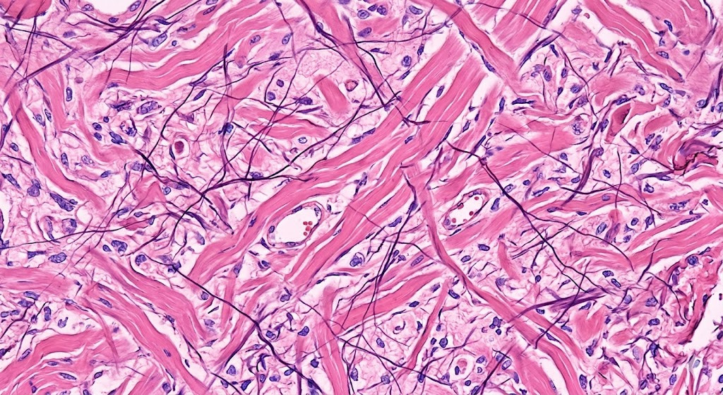
The extracellular matrix is composed of fibers and ground substance. This matrix determines the physical properties of the tissue. Some connective tissues are soft and flexible, while others are hard and rigid.
Connective tissue performs numerous vital functions including structural support, transportation of substances, storage of energy, defense against infection, tissue repair, and protection of organs.
Components of Connective Tissue
Connective tissue consists of three main components: cells, fibers, and ground substance.
The cells may include fibroblasts, macrophages, mast cells, plasma cells, adipocytes, and wandering leukocytes. Fibroblasts are the most common cells and are responsible for producing fibers and ground substance.
The fibers are mainly collagen fibers, elastic fibers, and reticular fibers. Collagen fibers provide tensile strength, elastic fibers allow stretching and recoil, and reticular fibers form delicate supporting networks.
The ground substance is an amorphous gel-like material that fills the spaces between cells and fibers. It facilitates diffusion of nutrients and wastes.
Classification of Connective Tissue
Connective tissue is broadly classified into connective tissue proper, supporting connective tissue, and fluid connective tissue.
Supporting Connective Tissue
Supporting connective tissue forms the structural framework of the body. It provides strength, shape, support, and protection to various organs. This category mainly includes cartilage and bone. These tissues are more specialized and possess a firmer extracellular matrix compared to connective tissue proper.
Cartilage
Cartilage is a specialized connective tissue characterized by a firm yet flexible matrix. It is softer than bone but stronger than ordinary connective tissue. The cells of cartilage are known as chondrocytes, and these cells are located within small cavities called lacunae. The matrix of cartilage contains collagen or elastic fibers embedded within a gel-like ground substance.
One of the important features of cartilage is that it lacks blood vessels and nerves. Nutrients reach the chondrocytes through diffusion from surrounding tissues. As a result, cartilage heals slowly after injury.
Cartilage provides flexibility and support to structures while also reducing friction at joints. It is essential during embryonic development because much of the skeleton is initially formed as cartilage before being replaced by bone.
Types of Cartilage
Cartilage is classified into three major types based on the nature of fibers present in the matrix: hyaline cartilage, elastic cartilage, and fibrocartilage.
Hyaline Cartilage
Hyaline cartilage is the most common type of cartilage found in the human body. It possesses a smooth, translucent, bluish-white appearance. The matrix contains fine collagen fibers that are not easily visible under the microscope.
Hyaline cartilage is found at the ends of long bones in movable joints, in the nose, larynx, trachea, bronchi, and costal cartilages of ribs. In developing embryos, it forms the temporary skeleton.
The smooth surface of hyaline cartilage reduces friction between bones during movement. It also acts as a shock absorber and provides flexible support to respiratory passages.
Elastic Cartilage
Elastic cartilage contains abundant elastic fibers within its matrix, making it highly flexible and resilient. The tissue can bend repeatedly without losing its original shape.
It is found in structures requiring flexibility such as the external ear, epiglottis, and auditory tube.
The major function of elastic cartilage is to maintain the shape of organs while permitting flexibility.
Fibrocartilage
Fibrocartilage is the strongest type of cartilage because it contains dense bundles of collagen fibers. It is capable of resisting both compression and tension.
This cartilage is found in intervertebral discs, pubic symphysis, and menisci of the knee joint.
Fibrocartilage acts as an excellent shock absorber and provides mechanical support in areas subjected to heavy pressure.
Bone
Bone is a highly specialized, hard connective tissue that forms the skeletal framework of the body. Unlike cartilage, bone possesses a rigid mineralized matrix rich in calcium phosphate and collagen fibers. Bone tissue provides support, protection, movement, mineral storage, and blood cell formation.
Bone is a living tissue supplied with blood vessels and nerves. It undergoes continuous remodeling throughout life.
The cells of bone include osteoblasts, osteocytes, and osteoclasts. Osteoblasts are bone-forming cells, osteocytes are mature bone cells, and osteoclasts are involved in bone resorption.
Structure of Bone
Bone consists of an organic matrix containing collagen fibers and an inorganic component made of mineral salts, mainly calcium phosphate. The hardness of bone is due to mineralization.
Microscopically, compact bone contains cylindrical structures called osteons or Haversian systems. Each osteon consists of concentric layers known as lamellae surrounding a central canal that contains blood vessels and nerves.
Spongy bone contains irregular spaces filled with bone marrow. Red bone marrow produces blood cells, whereas yellow marrow stores fat.
Location of Bone
Bones form the entire skeleton of the body including:
- Skull
- Vertebral column
- Ribs
- Limbs
- Pelvis
Functions of Bone
Bone performs numerous essential functions. It provides structural support and maintains body shape. Bones protect delicate internal organs such as the brain, heart, and lungs. Along with muscles, bones enable body movement. Bone tissue also serves as a reservoir for calcium and phosphorus. Additionally, red bone marrow is the site of hematopoiesis, the process of blood cell formation.
Fluid Connective Tissue
Fluid connective tissues are specialized connective tissues with a liquid matrix. They are involved mainly in transportation and defense.
The two main fluid connective tissues are blood and lymph.
Blood
Structure of Blood
Blood is a fluid connective tissue composed of plasma and formed elements. Plasma is the liquid portion containing water, proteins, electrolytes, nutrients, hormones, and waste products.
The formed elements include:
- Red blood cells (erythrocytes)
- White blood cells (leukocytes)
- Platelets (thrombocytes)
Red blood cells contain hemoglobin and transport oxygen. White blood cells participate in immunity and defense. Platelets help in blood clotting.
Location of Blood
Blood circulates throughout the cardiovascular system within arteries, veins, capillaries, and the chambers of the heart.
Functions of Blood
Blood transports oxygen, nutrients, hormones, and waste products throughout the body. It helps regulate body temperature, pH, and fluid balance. White blood cells protect the body against infections, while platelets prevent excessive blood loss by clot formation.
Lymph
Structure and Location
Lymph is a clear, colorless fluid derived from tissue fluid. It circulates through lymphatic vessels and lymph nodes.
Functions of Lymph
Lymph returns excess tissue fluid to the bloodstream, transports absorbed fats from the intestine, and plays a crucial role in immunity through lymphocytes.
C. Muscular Tissue
Muscular tissue is specialized for contraction and movement. It is composed of elongated cells called muscle fibers that contain contractile proteins responsible for generating force. Muscular tissue enables body movement, maintenance of posture, heat production, and movement of substances within the body.
Muscle cells possess the unique property of excitability and contractility. When stimulated, they shorten and produce movement.
Muscular tissue is classified into three types:
- Skeletal muscle
- Smooth muscle
- Cardiac muscle
Each type differs in structure, location, control, and function.
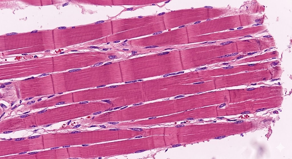
Skeletal Muscle
Structure of Skeletal Muscle
Skeletal muscle consists of long, cylindrical, multinucleated fibers arranged parallel to each other. The fibers show alternating light and dark bands known as striations, giving the tissue a striped appearance.
The nuclei are located at the periphery of the fibers. Skeletal muscles are voluntary muscles, meaning their activity is under conscious control.
The contractile proteins actin and myosin are organized into repeating units called sarcomeres, which are responsible for striations and contraction.
Location of Skeletal Muscle
Skeletal muscles are attached to bones by tendons. Examples include:
- Biceps
- Triceps
- Quadriceps
- Muscles of face
- Diaphragm
Functions of Skeletal Muscle
Skeletal muscles produce voluntary movements such as walking, writing, speaking, and facial expressions. They maintain posture and stabilize joints. Muscle contractions also generate heat, helping maintain body temperature.
Smooth Muscle
Structure of Smooth Muscle
Smooth muscle consists of spindle-shaped cells with a single centrally located nucleus. Unlike skeletal muscle, smooth muscle fibers do not show striations because the contractile proteins are arranged irregularly.
Smooth muscles are involuntary and function automatically under control of the autonomic nervous system and hormones.
Location of Smooth Muscle
Smooth muscle is present in the walls of hollow organs including:
- Stomach
- Intestines
- Blood vessels
- Urinary bladder
- Uterus
- Respiratory passages
Functions of Smooth Muscle
Smooth muscle contractions help propel substances through internal organs. In the digestive tract, rhythmic contractions known as peristalsis move food. In blood vessels, smooth muscles regulate blood pressure by controlling vessel diameter. In the uterus, smooth muscle contractions aid childbirth.
Cardiac Muscle
Structure of Cardiac Muscle
Cardiac muscle is found only in the heart. It consists of short, branched, striated fibers connected by specialized junctions called intercalated discs.
The cells usually contain one centrally located nucleus. Cardiac muscle is involuntary and possesses automatic rhythmic contractions.
Intercalated discs allow rapid transmission of impulses, enabling synchronized contraction of the heart.
Location of Cardiac Muscle
Cardiac muscle forms the muscular wall of the heart known as the myocardium.
Functions of Cardiac Muscle
The rhythmic contractions of cardiac muscle pump blood throughout the body continuously without fatigue. This ensures proper circulation of oxygen, nutrients, hormones, and waste products.
D. Nervous Tissue
Nervous tissue is specialized for communication and coordination. It forms the nervous system, which controls and integrates body activities. Nervous tissue can detect stimuli, generate electrical impulses, and transmit information rapidly throughout the body.
This tissue is highly specialized and responsible for functions such as sensation, memory, thinking, emotions, reflexes, and voluntary movement.
Nervous tissue consists of two major types of cells:
- Neurons
- Neuroglial cells
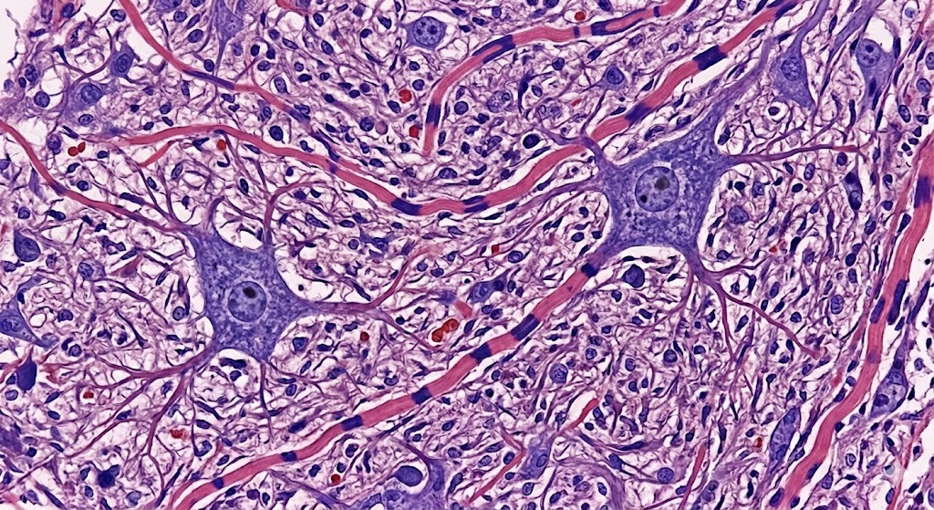
Neurons
Structure of Neurons
Neurons are the structural and functional units of the nervous system. Each neuron consists of three main parts:
- Cell body
- Dendrites
- Axon
The cell body contains the nucleus and cytoplasm. Dendrites are short branching processes that receive signals from other neurons. The axon is a long process that conducts impulses away from the cell body.
Some axons are covered by a myelin sheath, which increases the speed of impulse conduction.
Location of Neurons
Neurons are present in:
- Brain
- Spinal cord
- Peripheral nerves
- Sensory organs
Functions of Neurons
Neurons receive stimuli, process information, and transmit impulses to other neurons, muscles, or glands. They form complex communication networks responsible for body coordination and responses.
Neuroglial Cells
Structure and Role
Neuroglial cells, or glia, are supporting cells of nervous tissue. Unlike neurons, they do not conduct impulses. Instead, they provide nourishment, protection, insulation, and support to neurons.
Different types of glial cells perform specialized functions such as forming myelin, removing debris, and maintaining the chemical environment around neurons.
Functions of Nervous Tissue
Nervous tissue performs several critical functions in the body. It detects changes in both internal and external environments, conducts impulses rapidly, coordinates muscular activity, regulates glandular secretions, maintains homeostasis, and enables higher mental functions such as learning, memory, reasoning, and emotions.
Conclusion
The human body is composed of four fundamental tissues: epithelial, connective, muscular, and nervous tissues. Each tissue possesses unique structural characteristics that directly relate to its specialized functions. Epithelial tissue protects and lines body surfaces, connective tissue supports and binds structures, muscular tissue enables movement through contraction, and nervous tissue coordinates body activities through impulse transmission.
Together, these tissues work harmoniously to maintain the structure, function, and survival of the body. Understanding tissue organization is essential for comprehending normal physiology, pathology, and medical sciences.