
Aim: Identification of Axial Bones
References:
- Structural Organisation In Animals-Animal Tissues, Trueman’s Elementary Biology, K.N.
Bhatia, et al, Edition 2016, pp. 168-213
- Haematology, Practical Human Anatomy and Physiology, S.R. Kale et al., Nirali Prakashan,
Eighth Edition, 2002, pp. 5-9
Theory:
The axial skeleton is one of the two main divisions of the human skeleton, the other being the
appendicular skeleton. The axial skeleton forms the central axis of the body. It consists of bones
that provide support, protection, and the framework for various vital structures, such as the
brain, spinal cord, and organs in the thoracic cavity. It includes 80 bones which are as follows:
Skull
The skeleton of the head is called the skull. It rests upon the upper end of the vertebral column.
Its bony structure consists of the following parts:
Bones of Cranium: The cranium is formed by 8 bones. The bones that form the cranium are:
1 frontal bone: The bone of the forehead, involved in the formation of orbital cavities and the
prominent ridges above the eye.
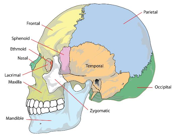
2 parietal bones: Forming the sides and roof of the skull.
2 temporal bones: Located on each side of the head.
1 occipital bone: Forming the back of the head.
1 sphenoid bone: A bat-shaped bone with its wings outstretched, occupying the middle
portion of the base of the skull.
1 ethmoid bone: Occupying the anterior part of the base of the skull.

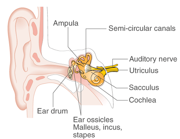
Ear Ossicles: There are 6 ear ossicles in the skull:
2 Malleus: The malleus, also known as the “hammer,” is one of the three ossicles in the middle
ear. It connects the eardrum to the incus, transmitting sound vibrations from the eardrum to the
inner ear, where they are converted into nerve signals for the brain to interpret.
2 Incus: The incus, or “anvil,” is one of the three ossicles in the middle ear. Positioned between
the malleus and the stapes, it receives sound vibrations from the malleus and transfers them to
the stapes. This relay amplifies sound waves, enabling effective transmission to the inner ear,
where they are converted into nerve signals for the brain to process as sound.
2 Stapes: The stapes, or “stirrup,” is the smallest bone in the body and the final ossicle in the
middle ear. It connects the incus to the cochlea’s oval window, transmitting sound vibrations to
the inner ear, where they are converted into nerve signals for the brain to interpret as sound.
Hyoid Bone: A single horseshoe-shaped bone providing support and protection to the throat.
Bones of the Face: 14 bones form the skeleton of the face:
2 zygomatic bones: Forming the cheeks.
2 maxilla: Forming the upper jaw.
2 nasal bones: Forming the lateral and superior surfaces of the bridge of the nose.
2 lacrimal bones: Located posteriorly and laterally to the nasal bones.
1 vomer: A thin flat bone extending upward from the middle of the hard palate, separating
the two nasal cavities.
2 palatine bones: “L” shaped bones forming the posterior part of the hard palate.
2 inferior nasal conchae: Forming the lateral part of the nasal cavity.
1 mandible: One of the strongest bones of the body, and the only movable bone of the skull.
Vertebral Column (Backbone)
The vertebral column is about 71 cm long and lies in the mid-dorsal line of the neck and trunk.
It is made up of 33 vertebrae, grouped into 5 categories:
Cervical Vertebrae: 7 in number, present in the neck. The first cervical vertebra is called the
atlas, and the second is called the axis.

Thoracic Vertebrae: 12 in number, present in the chest. They are larger and stronger than the
cervical vertebrae.
Lumbar Vertebrae: 5 in number, present in the abdomen. They are the largest and strongest
in the vertebral column.
Sacrum: 5 sacral vertebrae are fused in adults to form a single structure called the sacrum.
Coccyx: The four coccygeal vertebrae are fused to form a curved triangular bone called the
coccyx, considered a vestigial tail.
Sternum (Breast Bone)
A flat bone present in the chest, about 15 cm long. It consists of three parts:
Manubrium: The uppermost part with articular facets laterally for articulation with the
clavicle to form the sternoclavicular joint.
Body: The middle portion, which has facets for articulation with ribs.
Xiphoid Process: The tip of the bone.
Ribs
Twelve pairs of ribs form the bony lateral walls of the thoracic cage.
True Ribs: The first seven pairs are called true ribs because their anterior ends are attached
directly to the sternum by means of small pieces of cartilage.
False Ribs: The eighth, ninth, and tenth pairs of ribs are called false ribs. They articulate by
cartilage with the costal cartilage of the seventh rib and thus are attached indirectly to the
sternum.
Floating Ribs: The last two pairs of ribs are called floating ribs because their anterior ends are
not attached to either the sternum or the cartilage of another rib.
Each rib is a flat bone with a head (articulates with the body of vertebra), neck, tubercle
(articulates with thoracic vertebra), and angle (the point at which the bone ends).
🚀 Don’t miss out on this opportunity to supercharge your education. Download now and embark on a journey of continuous growth and knowledge!
Happy Learning,
pharmaacademias.com Team