
Aim- Identification of appendicular bones.
Reference:
- Structural Organisation In Animals-Animal Tissues, Trueman’s Elementary Biology, K.N.
Bhatia, et al., Edition 2016, pp. 168-213 - Haematology, Practical Human Anatomy And Physiology, S.R. Kale et al., Nirali Prakashan,
Eight Edition, 2002, pp. 5-9.
Theory
It is situated at the lateral side, which extends outwards from the principal axis. The
appendicular skeleton system comprises the pectoral girdle (shoulder girdle), pelvic girdle (hip
girdle), upper limbs or arms bones, and lower limbs or legs.
PECTORAL GIRDLE
Each pectoral girdle consists of two bones: 1 clavicle and 1 scapula.
Scapula- The scapula is a flat bone with a ridge called the spine and a triangular body. The
spine ends in an expanded part called the acromion, which connects to the clavicle. On the
lateral end, there’s a projection called the coracoid process, which attaches to muscles and
ligaments. The scapula’s body has two surfaces.
i) The costal surface, which is concave and marked by ridges.
ii) The dorsal surface, divided into two parts by the spine into upper small
supraspinous fossa and lower area forming infraspinous fossa.
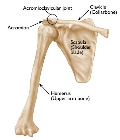
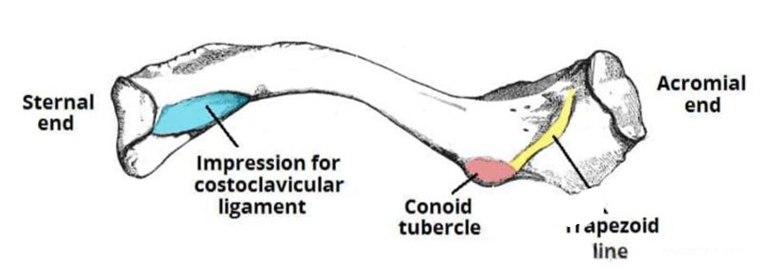
Clavicle: The collar bone, or clavicle, connects the scapula to the sternum. It’s “S”-shaped,
with a shaft and two ends: acromial (flat) and sternal (quadrangular).
BONES OF THE ARMS
Each arm consists of 30 bones: 1 humerus, 1 radius, 1 ulna, 8 carpal bones, 5 metacarpal bones,
5 digits (14 phalanges).
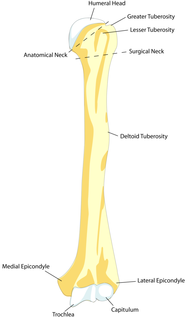
Humerus: The humerus, the longest and strongest upper arm bone, has a proximal, shaft, and
distal end. The proximal end includes the head, neck, and lower tubercle. The distal end has
the capitulum, trochlea, and fossae.
Ulna: The ulna, found on the inner side of the forearm, is longer than the radius. Its proximal
end includes the olecranon and coronoid processes, while the distal end has a smooth surface
for joining with the radius and a styloid process for ligaments.
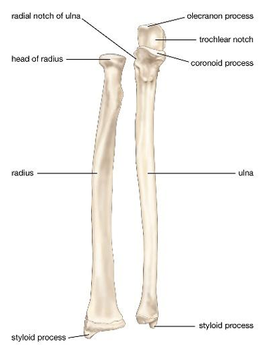
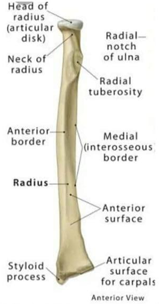
Radius: The radius, a forearm bone, has a proximal, shaft, and distal end. Its upper end has a
disc-shaped head that connects with the humerus’s capitulum and a narrow neck below it. The
distal end widens for articulation with carpal bones.
Carpal, metacarpal, and phalanges: The wrist comprises two rows of eight carpals: scaphoid,
lunate, triquetrum, and pisiform in the proximal row, and trapezium, trapezoid, capitate, and
hamate in the distal row. The palm is formed by five metacarpals, each with a proximal end
connecting to carpals, a middle shaft, and a distal end linking to phalanges. Fourteen phalanges
make up the fingers and thumb, with each bone called a phalanx.
PELVIC GIRDLE
The pelvis comprises two hip bones, each consisting of three separate bones: the ilium, ischium,
and pubis. The acetabulum forms the hip joint, a deep depression on the outer surface
connecting with the femur’s head. The ileum is a flat plate above the acetabulum, featuring the iliac crest on its upper border and a greater sciatic notch. The ischium is below and behind the
acetabulum, while the pubis is in front and below it. The ischium has a lesser sciatic notch, and
there’s an opening called the obturator foramen.
BONES OF THE LEGS
Each leg consists of 30 bones: 1 femur, 1 tibia, 1 fibula, 1patella (knee cap), 7 tarsal bones, 5
metatarsal bones, 5 digits (14 phalanges).
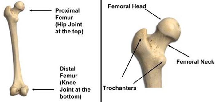
Femur: The femur, the body’s longest and strongest bone, has a rounded head at the upper end,
a constricted neck, and greater and lesser trochanters. The head connects to the pelvic girdle’s
acetabulum, and the neck leads to the long shaft with the trochanters. At the lower end are two
condyles, lateral and medial, separated by an intercondylar fossa.
Patella: It is a flat, sesamoid bone. It is roughly triangular.
Tibia: The second longest bone, is thicker and positioned more towards the front and inner
side of the leg. It has a broad upper end with two condyles and a flat lower end that forms the
ankle joint with the talus bone.
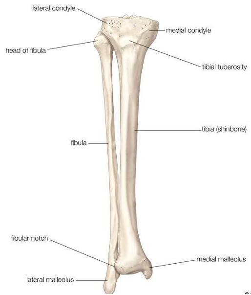
Fibula: The fibula is shorter, thinner, and positioned more laterally and deeply. Its head
articulates with the lateral condyle of the tibia, and the lower end connects to the lower part of
the tibia.
Tarsal, metatarsal and phalanges: The foot consists of seven tarsal bones (calcaneum, talus,
cuboid, navicular, and three cuneiforms), five metatarsal bones, and 14 phalanges—three in
each toe and two in each big toe.
🚀 Don’t miss out on this opportunity to supercharge your education. Download now and embark on a journey of continuous growth and knowledge!
Happy Learning,
pharmaacademias.com Team