


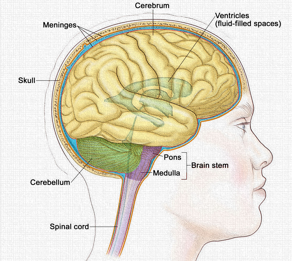
The cerebrum represents the largest and most highly developed part of the human brain. It is the seat of higher cognitive functions, voluntary motor control, sensory perception, language, reasoning, memory, and emotion. Anatomically, it forms the superior portion of the brain and accounts for nearly 80% of its total weight. Functionally, the cerebrum integrates complex neural processes that define consciousness, intelligence, personality, and behavior—attributes that distinguish humans from other species.
1. Anatomy of the Cerebrum
The cerebrum exhibits a highly organized and complex structure designed to maximize neural processing and intercommunication.
A. Gross Anatomy
- Cerebral Hemispheres:
The cerebrum is divided into two symmetrical halves, known as the right and left cerebral hemispheres, separated by a deep groove called the longitudinal fissure. Despite appearing symmetrical, each hemisphere exhibits functional specialization, a phenomenon known as lateralization. The corpus callosum, a thick band of myelinated nerve fibers, connects the two hemispheres and facilitates communication between them.

2. Surface Features:
The surface of the cerebrum is highly convoluted, consisting of gyri (ridges) and sulci (shallow grooves). This convoluted arrangement increases the surface area, allowing for a greater number of neurons and enhanced processing power within the limited cranial space.
- Fissures are deeper grooves, such as the central sulcus, lateral fissure (Sylvian fissure), and parieto-occipital sulcus, which divide the cerebrum into distinct lobes.
3. Cerebral Lobes:
Each hemisphere is divided into four primary lobes (frontal, parietal, temporal, and occipital) and a fifth, hidden lobe known as the insula. Each lobe performs specialized functions but works in harmony with others for integrated brain activity.
- Frontal Lobe: Involved in voluntary movement, reasoning, planning, problem-solving, and emotional regulation.
- Parietal Lobe: Processes sensory information such as touch, temperature, pain, and spatial orientation.
- Temporal Lobe: Associated with hearing, memory formation, and language comprehension.
- Occipital Lobe: Responsible for visual processing.
- Insula (Island of Reil): Plays a role in visceral sensation, emotional awareness, and autonomic control.
4. Gray and White Matter:
The cerebral cortex forms the outer layer of gray matter, consisting of neuron cell bodies, dendrites, and synapses. Beneath it lies the white matter, composed primarily of myelinated axons that form tracts connecting various brain regions.
- Gray Matter: Site of information processing, integration, and decision-making.
- White Matter: Responsible for rapid communication between cortical and subcortical areas.
5. Basal Nuclei (Basal Ganglia):
Deep within the cerebral hemispheres lie clusters of gray matter known as the basal nuclei (including the caudate nucleus, putamen, and globus pallidus). These structures are vital for motor coordination, habit formation, and regulation of voluntary movements.
6. Limbic System:
A complex network within the cerebrum that includes structures such as the hippocampus, amygdala, and cingulate gyrus. It governs emotional responses, motivation, and memory formation—linking emotion with cognition.
2. Physiology of the Cerebrum
The physiological functions of the cerebrum are diverse and interdependent, encompassing sensory reception, motor control, language processing, memory storage, and cognitive functions.
A. Sensory Functions
- The cerebrum interprets all sensory data received from the body via the thalamus.
- Specific regions of the cerebral cortex are designated as primary sensory areas, responsible for processing inputs such as touch, pressure, temperature, vision, hearing, taste, and smell.
- The primary somatosensory cortex (postcentral gyrus of the parietal lobe) receives information from sensory receptors distributed throughout the body, allowing perception of tactile sensations and proprioception.
B. Motor Functions
- The primary motor cortex (precentral gyrus of the frontal lobe) initiates voluntary muscle contractions.
- Adjacent areas like the premotor cortex and supplementary motor area coordinate complex, learned motor activities such as writing or playing a musical instrument.
- The basal nuclei and cerebellum work in coordination with the motor cortex to ensure smooth and precise movements, suppressing unwanted muscle activity.
C. Cognitive and Integrative Functions
- The cerebrum is the center of intellectual functions, including thought, reasoning, problem-solving, and judgment.
- It enables humans to perform abstract thinking, decision-making, and moral reasoning.
- Association areas within each lobe integrate sensory and motor information, forming complex patterns of perception, learning, and memory.
D. Language and Communication
- Language involves both comprehension and expression and is primarily localized in the left hemisphere of the cerebrum in most individuals.
- Broca’s area (frontal lobe) controls motor aspects of speech—formulating words and articulation.
- Wernicke’s area (temporal and parietal lobe junction) is essential for understanding spoken and written language.
- Coordination between these areas through the arcuate fasciculus allows fluent communication.
E. Memory and Learning
- Short-term memory is associated with transient electrical activity in the prefrontal cortex, while long-term memory involves structural changes in neuronal connections, particularly within the hippocampus and temporal lobe.
- Learning is facilitated by synaptic plasticity, where repeated stimulation strengthens synaptic connections—a process underlying long-term potentiation (LTP).
F. Emotional Regulation
- The limbic system and prefrontal cortex work together to process and regulate emotions.
- The amygdala is particularly involved in fear and aggression responses, whereas the hippocampus links emotions with memory formation.
- These emotional centers influence behavior, motivation, and decision-making.
3. Functional Areas of the Cerebrum
The cerebral cortex is functionally divided into three categories of cortical areas: sensory areas, motor areas, and association areas. Each region has a specialized role but operates in concert for integrated brain function.
A. Sensory Areas
- Primary Somatosensory Cortex (Postcentral gyrus – Parietal lobe): Receives input from receptors in the skin, muscles, and joints to interpret touch, temperature, pressure, and pain.
- Visual Cortex (Occipital lobe): Processes visual stimuli, including color, shape, and movement.
- Auditory Cortex (Temporal lobe): Interprets sound patterns and pitch.
- Olfactory Cortex (Temporal lobe): Responsible for the perception of smell.
- Gustatory Cortex (Insula and Frontal lobe): Interprets taste sensations.
B. Motor Areas
- Primary Motor Cortex (Precentral gyrus – Frontal lobe): Initiates voluntary skeletal muscle movement.
- Premotor Cortex: Plans and coordinates learned or complex movements.
- Frontal Eye Field: Controls voluntary eye movements.
- Broca’s Area: Controls the muscles involved in speech production.
C. Association Areas
These areas integrate information from multiple sensory inputs and link them to memories, emotions, and motor responses.
- Prefrontal Cortex: The “executive center” responsible for reasoning, planning, personality expression, and decision-making.
- Wernicke’s Area: Critical for language comprehension.
- Parietal-Temporal-Occipital Association Area: Integrates visual, auditory, and somatosensory data for complex perception and spatial awareness.
4. Clinical Correlations
- Damage to Broca’s area results in expressive aphasia (inability to speak fluently).
- Lesions in Wernicke’s area cause receptive aphasia (inability to understand language).
- Parietal lobe injury may lead to loss of spatial orientation or sensory neglect.
- Frontal lobe damage often results in personality changes and impaired judgment.
- Occipital lobe lesions may lead to partial or complete blindness.
Conclusion
The cerebrum is a masterpiece of biological architecture and function. Its elaborate structure, from gyri and sulci to interconnected networks of gray and white matter, allows humans to think, feel, perceive, and act with remarkable precision. Each lobe and cortical area contributes uniquely to the orchestration of life’s most intricate processes—memory, emotion, intelligence, and consciousness. Collectively, the cerebrum stands as the command center of human identity and intellect, defining the very essence of what it means to be sentient.